The Body's Lie About Where It Hurts
A heart attack often screams in the arm. The reason reveals how the brain builds pain from guesswork.
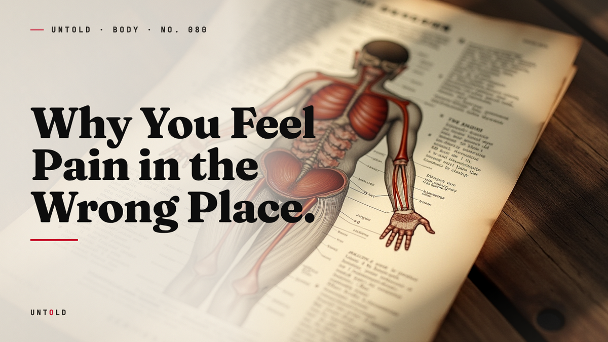
A man in his sixties sets down his coffee and rubs his left arm. It aches, a dull heaviness spreading toward the jaw. He assumes he slept on it wrong. Within the hour he is in an emergency room, and the electrocardiogram tells a story his arm could not: his heart is starving for oxygen. The muscle at the center of his chest is dying. Yet the loudest complaint came from a limb that was, anatomically speaking, perfectly fine.
This is one of the strangest facts about the human body, and one of the most consequential. Pain, which we treat as the most trustworthy of sensations, routinely lies about its own address. A diseased gallbladder can ache between the shoulder blades. A kidney stone can burn a searing line down into the groin. Eat ice cream too quickly and a spike of pain detonates behind your forehead, though nothing has touched your forehead at all. Physicians have a clinical name for this phenomenon: referred pain, agony experienced at a distance from its true source.
The puzzle is not merely academic. For more than a century, understanding why the body misreports its own injuries has been a matter of life and death. And the answer, buried in the crowded wiring of the spinal cord, overturns a comfortable assumption we all carry: that pain is a simple message, delivered faithfully from the site of harm to a passive brain waiting to receive it. It is nothing of the kind.
A map drawn on the skin
Long before anyone could explain referred pain, careful clinicians could not help but notice it. Patients with angina, the crushing chest discomfort of a failing coronary supply, kept describing pain that radiated into the left arm, the neck, the jaw. Patients with disease deep in the abdomen pointed instead to patches of skin that were themselves healthy. The pattern was too consistent to be coincidence, and too baffling to ignore.
The man who turned these scattered observations into a system was a British neurologist named Henry Head. In a landmark series of papers published from 1893 onward, Head charted the surface of the body against the organs beneath it, documenting the zones of skin that grew tender or painful when a particular internal organ was diseased.1 These regions came to be called Head’s zones, and versions of his diagrams still appear in medical textbooks today. Each internal organ, Head found, was linked to a specific territory on the skin, as if the viscera had signed their complaints in a code written across the body’s outer surface.
Head’s work was extraordinary partly because of how he pursued it. In one famous experiment, he had a colleague sever a nerve in his own forearm so that he could document, over the years of its slow regeneration, exactly how sensation returned and how the boundaries of feeling were mapped.2 He was a man willing to make his own body the laboratory. But mapping a pattern is not the same as explaining it. Head could show you, with precision, that a bad heart tended to announce itself in the left arm. He could not yet say why the arm should suffer for the sins of the heart.
That question, why the brain should get its own body map so badly wrong, would take another half century to answer. And the answer, when it came, was less about anatomy than about the strange logic of how the brain decides what a sensation means.
The crowded switchboard
Descend into the spinal cord and the tidy picture of separate nerves for separate body parts begins to dissolve. Sensory fibers do not each run a private line to the brain. Instead, signals from vast and distant regions of the body funnel into shared relay stations, clusters of neurons in the spinal cord’s dorsal horn, where a single second-order neuron may receive input from many sources at once.
Here is the crucial detail. The nerve fibers carrying information from an internal organ frequently arrive at the very same spinal neurons that receive fibers from a patch of skin. A single relay cell might be listening simultaneously to the heart and to the skin of the left arm, or to the diaphragm and to the shoulder. The two streams of information converge onto one pathway, and from that point upward toward the brain, they travel together, indistinguishable. It is a switchboard with crossed wires, and the crossing is built into the architecture from birth.
In 1961 the American physiologist Thomas Cleland Ruch gave this arrangement its enduring name and its explanatory power. His convergence-projection theory proposed that because organ fibers and skin fibers converge onto a shared ascending pathway, the brain receiving the resulting signal simply cannot tell which source fired it.3 The message arrives stripped of its return address. And so the brain does what brains always do with ambiguous information: it guesses. It projects the pain outward onto the location it deems most likely.
The question then becomes: why does the brain guess the skin, when the real emergency is the organ? The answer is a matter of statistics accumulated over a lifetime.
Why the brain trusts the surface
Think about where pain has actually come from across the whole of your life. Skinned knees, burned fingertips, paper cuts, stubbed toes, sunburn, splinters. The skin is a relentless reporter, sending the brain painful feedback thousands upon thousands of times, each one accompanied by a clear visible cause. The brain learns, over decades, that when this particular relay neuron lights up, the culprit is almost certainly something happening at the surface.
Internal organs, by contrast, are nearly silent. A healthy liver, a healthy heart, a healthy kidney will go an entire lifetime without generating a single painful signal. The viscera have very few pain receptors to begin with, and those they have are tuned to stretching, ischemia, and inflammation rather than to the cutting and burning that dominate the skin’s experience. The brain, in other words, has spent a lifetime practicing how to read the skin and has almost no experience reading the organs.
So when a coronary artery finally clogs and the starving heart muscle fires its alarm up the shared pathway, the brain reaches for the only interpretation it has ever needed. It assumes the signal must be coming from the skin, because the skin is where such signals nearly always come from. It projects the emergency onto the arm, the jaw, the shoulder: the familiar map. This is why referred pain is not random. It follows the ancient developmental logic of which body parts happened to share a spinal segment with which organ, a logic laid down in the embryo and preserved into adulthood.
The deeper implication is philosophical as much as physiological. Pain is not a readout. The brain does not passively receive a location and display it. The brain interprets, actively and constantly, and referred pain is what happens when its interpretation, sensible on the whole, misfires in a specific case. Experience, built for the common situation, overrides accuracy in the rare one.
The clock inside appendicitis
Few conditions illustrate this drama more clearly than appendicitis, and few have caught out more physicians. The appendix sits low on the right side of the abdomen, yet the pain of an inflamed appendix classically begins nowhere near it. It starts as a vague, diffuse ache around the navel, poorly localized, hard for the patient to pin down.4
That early pain is referred. The appendix, like the rest of the midgut, sends its visceral signals into spinal segments that the brain associates with the region around the belly button, and so that is where the alarm first appears. It is dull and central precisely because visceral pain is imprecise by nature. Only hours later, once the inflammation has spread to the lining of the abdominal wall directly over the appendix, does the pain migrate and sharpen into the pointed lower-right tenderness that surgeons recognize. That later pain is not referred at all. It comes from somatic nerves in the abdominal wall itself, nerves that are excellent at localization.
The two-act structure is diagnostically vital and, when misread, dangerous. A meaningful fraction of patients present early, while the pain is still vague and central, and the misleading location has led to missed or delayed diagnoses. The migration of the pain over time is itself the clue: a moving target that tells the trained clinician which organ is speaking and how far its inflammation has progressed.
The fastest wrong answer
Not all referred pain unfolds over hours. Some of it is nearly instantaneous, and almost everyone has felt it. Bite into something frozen too fast and a stabbing pain erupts behind your forehead and temples within seconds. Doctors call it sphenopalatine ganglioneuralgia. Everyone else calls it brain freeze.
The roof of your mouth is nowhere near your forehead, and yet that is where the pain seems to land. In 2012 a research team led by Jorge Serrador set out to watch the phenomenon happen. By having volunteers sip ice water against the upper palate while their cerebral blood flow was monitored, the researchers documented a sudden surge of blood into the anterior cerebral artery, followed swiftly by the headache, and then a return to baseline as the vessel constricted again.5 The rapid dilation of a blood vessel appeared to be the trigger.
The cold stimulus lands on the palate, but the signal is carried by branches of the trigeminal nerve, the great sensory nerve of the face. That same nerve also serves the forehead. When it fires in response to the cold above, the brain, once again reading from experience, attributes the pain to the region the trigeminal nerve most often reports from: the front of the head. Same nerve, wrong reported location. Brain freeze is referred pain in miniature, a laboratory demonstration you can run with a milkshake.
Not a bug but a strategy
It is tempting to file all of this under the heading of error, a quirk of sloppy wiring that evolution never bothered to clean up. That framing is almost certainly wrong, and getting it wrong obscures the most interesting part of the story.
Consider the alternative. Imagine the heart could report its distress accurately, as a faint, diffuse ache somewhere deep and central in the chest, unlocatable and easy to dismiss. That kind of signal is nearly useless for survival. Vague internal aches do not make an animal flee a predator, protect a wound, or seek help. They are too easy to ignore. The skin’s pain, by contrast, is sharp, urgent, and precisely located, exactly the sort of alarm that produces immediate action.
Seen this way, referred pain looks less like a malfunction and more like a translation. The brain takes a signal it has no good vocabulary for, the silent complaint of an organ, and renders it in the one language it commands fluently: the vivid, actionable grammar of surface pain. The arm pain of a heart attack is not the heart making a mistake. It is the nervous system converting a quiet internal catastrophe into a loud, locatable emergency that a person cannot easily ignore. The very inaccuracy that misleads is what compels a response.
This reframing turns a bug into a feature. The system did not evolve to give physicians an accurate map. It evolved to keep the organism alive, and a loud alarm in the wrong place beats a silent one in the right place.
Reading the disguise
For the clinician, referred pain is a coded language, and learning to read it is a core skill of diagnosis. Jaw pain that arrives with exertion may be the heart. Right shoulder tip pain may be irritation of the diaphragm from bleeding or inflammation below it. Mid-back pain that will not ease may be the pancreas. Each of these patterns is a clue left by the shared wiring of the spinal cord, a fingerprint of which organ converges onto which segment.
This matters most in the cases where the disguise is most complete. A significant proportion of heart attacks are described as silent or atypical, their chest component muted or absent, their warning delivered instead as arm, jaw, back, or even abdominal discomfort that patients and sometimes doctors mistake for indigestion. Women, older adults, and people with diabetes are more likely to experience these atypical presentations, and the delay in recognizing them costs lives.6 The clinician who has internalized the map of referred pain is the one who asks the further question when the pain seems to sit in an unlikely spot.
The lesson for the rest of us is not to self-diagnose from a chart of Head’s zones. It is subtler than that. It is to abandon the intuition that pain is a reliable pointer, and to treat a strange or unaccountable pain, especially one accompanied by breathlessness, sweating, or a sense of dread, as worth taking seriously rather than rubbing away.
Coda
We trust pain because it feels like the most direct testimony the body can give, a raw fact rather than an opinion. Referred pain quietly dismantles that trust and replaces it with something more remarkable. Every pain you have ever felt was an interpretation, a best guess assembled by a brain working from limited information and a lifetime of statistics. Usually the guess is right, and the location it hands you is true. Occasionally, when a silent organ finally cries out, the guess is wrong, and the pain lands somewhere it was never born. But even then the error is a kind of wisdom. The body, unable to make you hear a whisper from within, has learned to shout in a language you cannot help but understand.
Sources
- Head, H., “On disturbances of sensation with especial reference to the pain of visceral disease,” Brain, 1893. — https://academic.oup.com/brain/article-abstract/16/1-2/1/253066
- Rivers, W. H. R. and Head, H., “A human experiment in nerve division,” Brain, 1908. — https://academic.oup.com/brain/article/31/3/323/263661
- Ruch, T. C., “Pathophysiology of Pain,” in Physiology and Biophysics (Ruch & Patton, eds.), W. B. Saunders, 1961. — https://www.ncbi.nlm.nih.gov/books/NBK553085/
- Craig, A. D., “Pain mechanisms: labeled lines versus convergence in central processing,” Annual Review of Neuroscience, 2003. — https://www.annualreviews.org/doi/10.1146/annurev.neuro.26.041002.131022
- Serrador, J. M. et al., “Cerebral vascular blood flow changes during ‘brain freeze,’” FASEB Journal / Experimental Biology, 2012. — https://www.sciencedaily.com/releases/2012/04/120422162536.htm
- Canto, J. G. et al., “Presenting Symptoms of Acute Myocardial Infarction and Their Association With Sex and Mortality,” JAMA, 2012. — https://jamanetwork.com/journals/jama/fullarticle/1103172
- Andersson, R. E., “The natural history and traditional management of appendicitis revisited,” World Journal of Surgery, 2007. — https://link.springer.com/article/10.1007/s00268-006-0056-y
Related reading