The Woman With a Y Chromosome in Her Brain
A century of biology said your body was one sealed genome. The autopsy table proved otherwise.
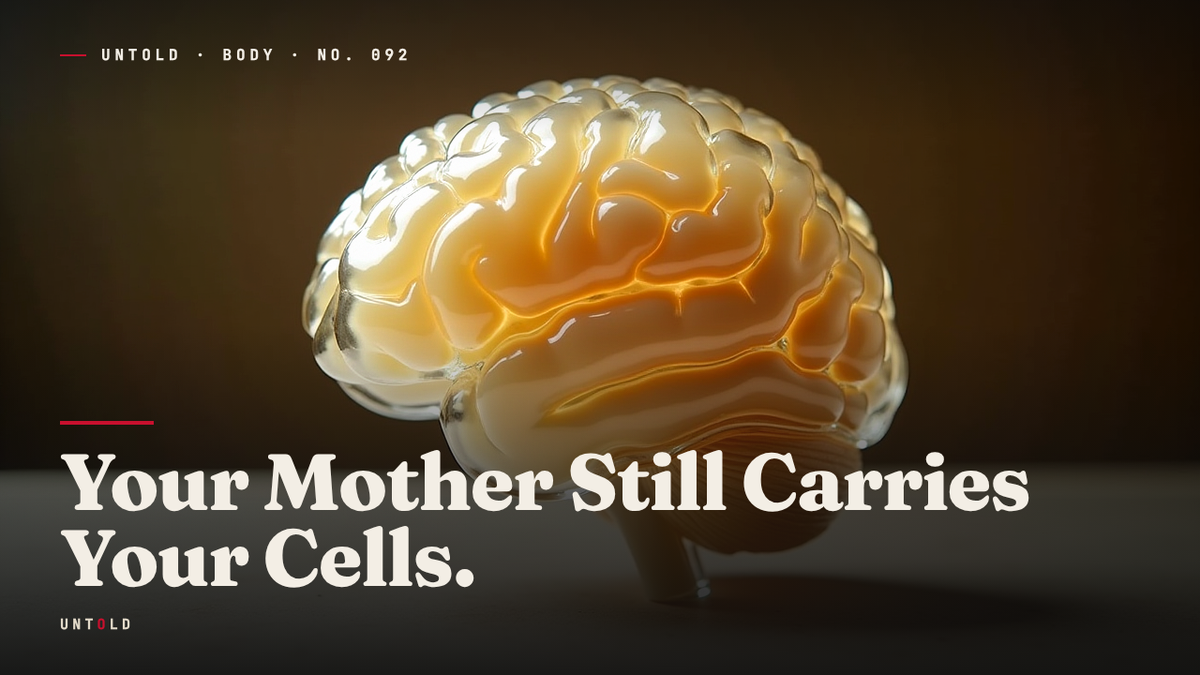
A pathologist draws a blade through a thin section of brain tissue. The woman on the table lived a long life and died in old age, and there is nothing remarkable about the specimen until it reaches the microscope. Then something appears that should not be there. A Y chromosome. Male DNA, unmistakable, threaded into the tissue of a woman who never possessed a Y chromosome of her own, who was female her entire life, who carried the standard set of two X chromosomes in every cell she was born with.
The DNA did not belong to her. It belonged to her son, born decades before she died. And somehow a piece of him had crossed into her body, migrated past one of the most tightly guarded barriers in human physiology, and lodged inside her brain, where it stayed for the rest of her life.
This is not a single strange case. When researchers examined the brains of fifty-nine deceased women, they found this male DNA in sixty-three percent of them.1 Nearly two out of every three women who had borne sons carried fragments of those sons inside their skulls, sometimes into their nineties, long after the pregnancies that produced them had faded into distant memory. The finding forced biology to confront a possibility it had spent a century denying: the human body is not one genome, sealed at birth. It is something stranger and more porous than that.
The One-Body Rule
For most of the twentieth century, biology taught a clean and reassuring idea about identity. One organism, one genome. Every cell in your body carries the same DNA, the same instructions, copied faithfully from the single fertilized egg you began as. Your genetic self was fixed at conception and sealed for life. Whatever else changed about you, the code did not.
Pregnancy seemed to fit neatly into this model. The placenta was understood as a perfect wall, a biological border checkpoint that let nutrients and oxygen pass between mother and child while keeping their two circulatory systems strictly apart. Two blood supplies. Two genomes. Never mixing. The fetus grew inside the mother, but it was walled off from her, its DNA quarantined behind the placental barrier. That separation was not just a convenience of textbook diagrams. It was thought to be a fundamental fact of mammalian reproduction.
The first crack in that wall appeared in 1893, though almost no one noticed. A German pathologist named Georg Schmorl was studying women who had died of eclampsia, the violent pregnancy complication that raises blood pressure to lethal levels. Examining their lungs under the microscope, he found something that did not belong: cells that appeared to have come from the placenta and the fetus, lodged deep in the maternal tissue.2 The cells had crossed the wall.
Schmorl assumed he was looking at an accident. These were fatal pregnancies, catastrophic and violent, and it seemed reasonable that the barrier might rupture under such extreme conditions, letting fetal cells leak through where they normally could not. He filed the observation as a curiosity of pathology, a byproduct of disaster rather than a feature of ordinary life. For decades, no one looked closer. The wall, everyone agreed, was still a wall. Schmorl had simply caught it breaking.
A Geneticist Runs the Numbers
The question stayed dormant until the 1990s, when a geneticist named Diana Bianchi decided to look for fetal cells not in the ruined bodies of women who had died in childbirth, but in the ordinary blood of healthy mothers going about their lives. If the barrier leaked only in catastrophe, she should find nothing. If it leaked routinely, she would find fetal cells circulating quietly in women who had delivered years or decades earlier.
The difficulty was distinguishing a mother’s cells from her child’s. A cell is a cell under the microscope, and maternal and fetal cells look identical. Bianchi needed a marker, a genetic signature that could belong only to the child. The Y chromosome gave her one. A woman does not carry a Y chromosome. So if male DNA turned up in the blood of a woman who had given birth to a son, there was only one plausible explanation. It had come from the son.
Bianchi sampled women who had delivered boys, some of them many years earlier, and searched their blood for the telltale male signature. She found it, and she found it far more often than the accident theory could explain. In her landmark 1996 study, published in the Proceedings of the National Academy of Sciences, she reported male fetal cells persisting in a mother’s blood as long as twenty-seven years after the birth of her son.3 Twenty-seven years. Long after the pregnancy ended, long after the child had grown into an adult, the cells were still there, circulating, surviving, replicating.
These were not visitors passing through. They had moved in. Bianchi gave the phenomenon a name that has stuck ever since: fetal microchimerism. The term borrows from the chimera of Greek myth, the creature stitched together from lion, goat, and serpent, a single body made of many animals. A mother, it turned out, was genetically more than one person. She carried a small resident population of her children’s cells inside her, a living minority genome woven into her own.
The implications rippled outward quickly. If fetal cells could survive in the blood for decades, where else did they go? Researchers began finding them everywhere they looked. In the thyroid. In the liver. In the skin. In the lungs, as Schmorl had glimpsed a century before, only now in living, healthy women rather than the dead. The cells did not stay confined to the bloodstream. They settled into organs and made themselves at home.
Into the Brain
Still, one boundary seemed too formidable to cross. The brain is protected by the blood-brain barrier, a dense lining of tightly joined cells that seals the circulation of the brain off from the rest of the body. It is one of the strictest checkpoints in human physiology, evolved to keep pathogens, toxins, and stray cells out of the most delicate organ we possess. Fetal cells were not supposed to be able to breach it. The brain, surely, was the one place the archive could not reach.
A team of researchers, including William Chan and J. Lee Nelson at the Fred Hutchinson Cancer Research Center, decided to test that assumption directly. They could not sample living brains, so they turned to autopsy tissue, examining the brains of fifty-nine women who had died and searching for the one signature only a son could leave behind: male DNA.1
The result overturned the last redoubt of the closed-body model. In sixty-three percent of the brains, they found it. Male DNA, distributed not in a single spot but across multiple regions of the brain, present in women who had died at every age. The oldest woman in whom they detected these cells was ninety-four years old.1 The cells had outlived the pregnancy by the better part of a century, surviving decades inside the very organ that was supposed to be inaccessible to them. The blood-brain barrier, like the placenta before it, had turned out to be a doorway rather than a wall.
The 2012 study, published in PLOS ONE, unsettled the field. It was one thing for fetal cells to loiter in the bloodstream or embed in an organ with a permeable supply. It was another for them to colonize the brain and remain there for a lifetime. The finding raised questions no one had thought to ask. What were these cells doing? Were they inert, or were they active participants in the life of the tissue around them?
Not Passive Passengers
Evidence accumulated that the cells were doing something. They did not distribute themselves at random. Again and again, researchers found fetal cells clustering in exactly the places where the body was under stress. They gathered at wounds. They appeared in scar tissue and in the healing incisions of caesarean sections, congregating at the site of injury as if summoned.4
More striking still, they turned up in damaged hearts. Studies found fetal cells integrated into a mother’s heart muscle after cardiac injury, embedded in the tissue near the site of damage.5 And some of these cells behaved unlike ordinary blood cells. They appeared to act like stem cells, undifferentiated and versatile, capable of becoming whatever the surrounding tissue required. Researchers documented fetal cells that had apparently transformed into heart muscle, into liver cells, even into neurons.5 The child’s cells were not merely surviving inside the mother. They seemed to be responding to her body’s needs, migrating toward injury and adopting the identity of the tissue they joined.
It was a seductive story. Pregnancy leaves behind a small standing repair crew, drawn from the child, that spends the mother’s remaining years mending the damage of age and injury. The cells that crossed into her when she was young stay on as a kind of biological reserve, ready to help.
The Cells Cut Both Ways
But the story is not that clean, and the honest version is more ambiguous. Fetal cells do not always help. The same population that seems to heal a wound in one context has been linked to harm in another.
Some studies associate fetal microchimerism with autoimmune disease, conditions in which the immune system turns against the body’s own tissue.6 The logic is uncomfortable but plausible: cells that are genetically foreign, even if only slightly, may provoke immune conflict, or may become targets themselves, dragging the surrounding tissue into a war it did not start. Certain autoimmune disorders are far more common in women than in men and often surface in the years after childbearing, a pattern that has led some researchers to suspect a link, though the evidence remains contested.
Yet other studies point the opposite direction. Fetal cells have been associated with protection against some cancers, possibly by patrolling for and attacking abnormal cells, or by aiding the tissue repair that keeps a tumor from taking hold.6 The same cells that might attack a healthy joint might also defend against a malignancy. The picture that emerges is not of a benevolent repair crew or a hostile invader, but of something genuinely double-edged, a resident population whose effects depend on context, timing, and the particular body it inhabits. Microchimerism is not a gift or a threat. It is both, and which face it shows may vary from woman to woman.
A Quiet Archive
The traffic, it turns out, runs both ways. Just as a child leaves cells behind in the mother, the mother leaves her own cells in the child. Maternal cells cross the placenta in the other direction and take up residence in the developing fetus, which means that you may still carry cells from your own mother inside you, decades after your birth, distributed through your tissues the way your cells are distributed through hers.7
And the archive is larger than two people. A mother who has carried more than one child may pass cells from an older sibling into a younger one, cells that lingered from a previous pregnancy and were still circulating when the next began. You may carry fragments of a brother or sister you never knew shared your body. The genome you think of as uniquely yours is populated by quiet minorities: your mother, your siblings, and in a mother’s case, every child she ever carried.
This is what the autopsy table revealed, one Y chromosome at a time. The body is not a single sealed genome, fixed at conception and closed to the world. It is a porous, populated thing, a living record of every exchange that pregnancy set in motion. The closed self was always a fiction, a tidy assumption that could not survive a closer look at the tissue.
When a mother says her child is part of her, she is not speaking in metaphor. Somewhere in her blood, her liver, perhaps her brain, cells that began as her child are still alive, still working, still hers and not hers at once. She carries them without knowing it, for the rest of her life. The wall was never a wall. It was a doorway, and everyone who passed through left something behind.
Sources
- Chan, W. F. N. et al., “Male Microchimerism in the Human Female Brain,” PLOS ONE, 2012. — https://journals.plos.org/plosone/article?id=10.1371/journal.pone.0045592
- Lapaire, O. et al., “Georg Schmorl on trophoblasts in the maternal circulation,” Placenta, 2007. — https://www.sciencedirect.com/science/article/abs/pii/S0143400406000737
- Bianchi, D. W. et al., “Male fetal progenitor cells persist in maternal blood for as long as 27 years postpartum,” PNAS, 1996. — https://www.pnas.org/doi/10.1073/pnas.93.2.705
- Mahmood, U. & O’Donoghue, K., “Microchimeric fetal cells play a role in maternal wound healing after pregnancy,” Chimerism, 2014. — https://www.tandfonline.com/doi/full/10.4161/chim.28746
- Kara, R. J. et al., “Fetal cells traffic to injured maternal myocardium and undergo cardiac regeneration,” Circulation Research, 2012. — https://www.ahajournals.org/doi/10.1161/CIRCRESAHA.111.249037
- Boddy, A. M. et al., “Fetal microchimerism and maternal health: A review and evolutionary analysis of cooperation and conflict beyond the womb,” BioEssays, 2015. — https://onlinelibrary.wiley.com/doi/10.1002/bies.201500059
- Maloney, S. et al., “Microchimerism of maternal origin persists into adult life,” Journal of Clinical Investigation, 1999. — https://www.jci.org/articles/view/6611
Related reading