The Organ That Refuses to Scar
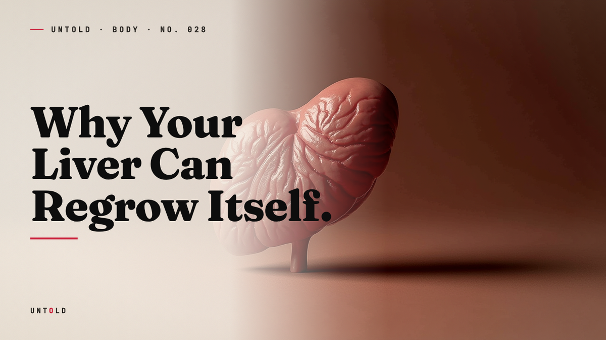
Cut away most of the liver and it rebuilds itself, but the way it does so is stranger than regrowth.
In 1931, two researchers at the University of Chicago performed an operation that sounds closer to vandalism than science. George Higgins and R. M. Anderson opened up an anesthetized rat, located its liver, and cut away roughly two thirds of the organ. Then they closed the animal up and waited.1
Under almost any other circumstance, removing two thirds of a vital organ is a death sentence. Take a third of a heart and the muscle that remains cannot grow to cover the loss; it scars, stiffens, and falters. Take a comparable share of a kidney or a lung and the organ simply makes do with less, forever diminished. But the rats in Higgins and Anderson’s lab did not falter. Within a week, the liver that remained had swelled back to almost exactly its original mass. The animals carried on as if nothing had been taken from them at all.
That experiment, dry and procedural in its published form, became the founding text of an entire field. Nearly a century later, the partial hepatectomy in rodents remains the standard laboratory model for studying how the liver rebuilds itself. And the central question Higgins and Anderson posed, almost by accident, has never fully been answered. How does a slab of tissue know what size it is supposed to be, grow back to precisely that size, and then stop?
A myth that anticipated the biology
The Greeks got there first, or at least they got somewhere close. In the myth of Prometheus, the Titan who stole fire from the gods and handed it to humankind, Zeus devises a punishment of exquisite cruelty. Prometheus is chained to a rock, and each day an eagle descends to feed on his liver. Each night the liver grows back, whole and ready, so that the torment can begin again at dawn. The agony is engineered to be eternal precisely because the organ renews itself.
It is tempting to read this as a lucky guess, a poet reaching for an image of endless suffering and landing, by chance, on the one organ in the body that actually does regenerate. But the choice may not have been arbitrary. Ancient anatomists, working with the bodies of wounded soldiers and sacrificial animals, were often startlingly accurate observers. The liver was central to early divination: Babylonian and Etruscan priests read the future in its lobes, which means they spent a great deal of time examining the organ closely. It is not impossible that someone, somewhere, noticed that a liver wound could heal in ways other wounds did not. Whether the Greeks encoded real observation into the Prometheus story or simply intuited something true, the myth lodged a fact in human memory thousands of years before anyone could explain it: the liver comes back.
What they could not have known is just how much the organ does, or how unusual its resilience is. The liver is the largest internal organ in the human body, weighing roughly three pounds in an adult. It sits in the upper right of the abdomen and functions less like a single organ than like an entire chemical plant running without a break. It filters toxins from the blood, breaks down drugs and alcohol, stores energy as glycogen, regulates blood sugar, manufactures the proteins that allow blood to clot, and produces the bile that lets the gut absorb fat. By most counts it performs over five hundred distinct metabolic tasks. A body can survive the loss of a kidney, a lung, a spleen, large stretches of intestine. It cannot survive without a liver for more than a day or two.
What actually grows back
Here the story takes a turn that confounds the intuitive picture, the one the Prometheus myth plants in the mind. When we imagine regeneration, we tend to picture a salamander regrowing a lost limb: a stump that sprouts, extends, and reconstitutes the missing shape piece by piece. That is not what the liver does.
Cut away two of the liver’s lobes and they do not come back. The missing tissue is gone for good, and the organ never returns to its original shape. What happens instead is that the cells in the remaining lobes go to work. They swell, and then they divide, all across the organ at once, until the total mass of the liver has been restored even though its form has not. The remaining lobes simply grow larger to make up the difference. Scientists distinguish this carefully from true regeneration and call it compensatory hyperplasia: a growth in cell number that compensates for what was lost rather than reconstructing it.
This distinction matters because it tells us where the work is being done. In most regenerating tissues, the heavy lifting falls to specialized stem cells, undifferentiated reserves that can become whatever the body needs. The liver mostly skips them. Its everyday workhorse cells, the hepatocytes that carry out all that metabolic labor, are themselves the agents of repair. Mature, fully differentiated, ordinarily content to sit quietly and filter blood, these cells abruptly re-enter the cell cycle and begin to divide. It is as though, after a fire, the office workers of a building put down their paperwork and rebuilt the walls themselves rather than waiting for a construction crew.
The speed of the response is extraordinary. Within minutes of injury, hundreds of genes that normally lie dormant in liver cells switch on. The organ that a moment earlier was a placid metabolic factory becomes a construction site, and it does so on a timescale measured in hours, not days.
The chemistry of an emergency
Much of what we understand about the molecular choreography of this process traces to the work of Nancy Fausto, a pathologist at the University of Washington who spent decades mapping the signals that wake the liver up. Her research, alongside that of George Michalopoulos and others, helped reveal a sequence that unfolds with the precision of an emergency protocol.2
It begins with priming. Two chemical messengers ring the first alarms. A signaling protein called interleukin-6, released in the wake of injury, rouses the resting hepatocytes from their quiescent state. Working in concert is tumor necrosis factor, or TNF, which primes those cells to begin dividing. Together these signals shift the liver cells from a state of rest into a state of readiness, like a fire crew pulling on their gear before the engine has even left the station.2
Then come the growth factors that actually drive proliferation. Chief among them is hepatocyte growth factor, which pushes the primed cells through the checkpoints of the cell cycle and into division. Epidermal growth factor and other signals reinforce the push. If interleukin-6 and TNF are the alarm bells, hepatocyte growth factor is the accelerator pressed to the floor. Under its influence, in the rodent models that Fausto and her colleagues studied so closely, an astonishing share of liver cells, well over half and by some measures more than seventy percent, begin dividing more or less simultaneously.2
And it is not only the hepatocytes that return. A liver is not a uniform block of identical cells but an intricately plumbed structure: a dense network of blood vessels, a branching system of bile ducts, supporting cells of several types, all arranged in a precise architecture. As the organ rebuilds its mass, this infrastructure reassembles too. The blood supply re-extends. The bile ducts re-form. The new tissue knits itself back into a functioning whole, not merely a heap of replacement cells.
The problem of knowing when to stop
For all the drama of the rebuilding, the deepest mystery in liver biology is not how the organ starts but how it knows to stop. The stakes of getting this wrong are severe in both directions. If the liver regrows too little, the body is left with insufficient function and slides toward organ failure. If it regrows too much, or fails to halt the frenzy of cell division once the work is done, it courts a different catastrophe: uncontrolled proliferation is the very definition of cancer. The liver must thread a needle. It has to restore itself to a target size and then, at exactly the right moment, apply the brakes.
Some of the most illuminating answers to how it manages this came not from the liver at all but from fruit flies. In the late 1990s and early 2000s, geneticists studying flies discovered a set of genes that, when mutated, caused tissues to overgrow grotesquely. One mutation produced flies with such excess tissue that researchers named the responsible gene after the bulkiest animal they could think of: the hippopotamus. The Hippo signaling pathway, as it came to be known, turned out to be one of biology’s master controls for organ size, conserved across species from insects to mammals.3
In the liver, the Hippo pathway functions as a kind of size sensor and emergency brake. When the organ is below its proper mass, the pathway permits growth, allowing a protein called YAP to drive cell proliferation. As the liver approaches its target size, Hippo signaling ramps up, restrains YAP, and tells the dividing cells to stand down. The construction crew puts away its tools. The hepatocytes that were dividing in a frenzy settle back into their quiet metabolic routine, and the organ returns to its watchful, filtering stillness. Disable the Hippo pathway in mice and their livers swell to several times the normal size, eventually developing tumors, a vivid demonstration of what the brake is for.3
Exactly how the liver senses that it has reached the right size remains incompletely understood. Mechanical cues, the physical crowding and tension between cells, the volume of blood flowing through the tissue, and chemical feedback all seem to play a part. The organ appears to hold, somewhere in the collective behavior of its cells, a memory of what it is supposed to be. It grows toward that target and halts when it arrives.
The limits of the power
The liver’s regenerative capacity is not a laboratory curiosity. Surgeons depend on it every day. Living-donor liver transplantation, now performed around the world, rests entirely on this biology. A healthy donor can give away a substantial portion of their liver, often the larger right lobe, to a patient whose own organ has failed. Within months, both the donor’s remaining liver and the transplanted portion in the recipient grow back to nearly full size. Two people walk away with functioning livers grown from what was once a single organ. No comparable operation is possible with the heart, the lungs, or the brain.
But the power has limits, and they are important to understand precisely because the organ’s resilience can lull us into treating it as indestructible. Liver regeneration works beautifully against acute insult: a single surgical resection, a discrete injury, a one-time toxic exposure. It copes far less well with chronic, repeated damage. Years of heavy alcohol use, persistent viral hepatitis, or the slow burn of fatty liver disease subject the organ to injury that never relents long enough for clean repair.
When damage is continuous, the liver’s response shifts in a way that ultimately undermines it. Instead of regenerating functional tissue, the organ lays down scar. Bands of fibrous, collagen-rich tissue accumulate, replacing working hepatocytes with inert connective material. This is cirrhosis, and it represents the failure of the very system that makes the liver remarkable. Scar tissue cannot divide. It cannot filter blood or build proteins or store energy. As it spreads, it chokes off the blood supply and crowds out the cells that could otherwise rebuild. At a certain point the regenerative machinery is simply overwhelmed, and the magic stops. The organ that could once recover from losing two thirds of itself can no longer recover at all.
This is the quiet warning folded into the biology. The liver is generous to a fault, and its generosity is finite.
Coda
The myth got the spirit of the thing right and the mechanism entirely wrong, which is perhaps the most a poet could hope for. The liver does not sprout back like Prometheus’s nightly regrowth, lobe restored to lobe in its original form. It does something less cinematic and more astonishing: its ordinary working cells, the ones quietly filtering your blood as you read this sentence, can drop their daily labor, multiply in their millions, rebuild a vascular and ductal architecture from the inside out, and then sense the exact moment to stop and resume their watch. They hold the organ’s correct size in some distributed memory and grow back toward it. The Greeks chained their hero to a rock and imagined eternal renewal. The truth is stranger and quieter, running every day inside a three-pound organ that asks for nothing and, until it is pushed past the edge of its patience, almost never quits.

Sources
- Higgins, G. M. & Anderson, R. M., Experimental pathology of the liver: restoration of the liver of the white rat following partial surgical removal, Archives of Pathology, 1931. — https://www.scinapse.io/papers/2417089983
- Fausto, N., Campbell, J. S. & Riehle, K. J., Liver regeneration, Hepatology, 2006. — https://pubmed.ncbi.nlm.nih.gov/16447274/
- Yu, F. X., Zhao, B. & Guan, K. L., Hippo Pathway in Organ Size Control, Tissue Homeostasis, and Cancer, Cell, 2015. — https://pubmed.ncbi.nlm.nih.gov/26544935/
- Michalopoulos, G. K. & DeFrances, M. C., Liver Regeneration, Science, 1997. — https://pubmed.ncbi.nlm.nih.gov/9082986/
- Taub, R., Liver regeneration: from myth to mechanism, Nature Reviews Molecular Cell Biology, 2004. — https://pubmed.ncbi.nlm.nih.gov/15459664/
- Pawlik, T. M. et al., Living Donor Liver Transplantation, surgical outcomes review, Liver Transplantation, 2005. — https://pubmed.ncbi.nlm.nih.gov/16035063/
- Power, C. & Rasko, J. E., Whither Prometheus’ Liver? Greek Myth and the Science of Regeneration, Annals of Internal Medicine, 2008. — https://pubmed.ncbi.nlm.nih.gov/18936507/
Related reading