The Language Your Skeleton Refuses to Hear
Bone only rebuilds when a load surprises it, and a daily walk never does.
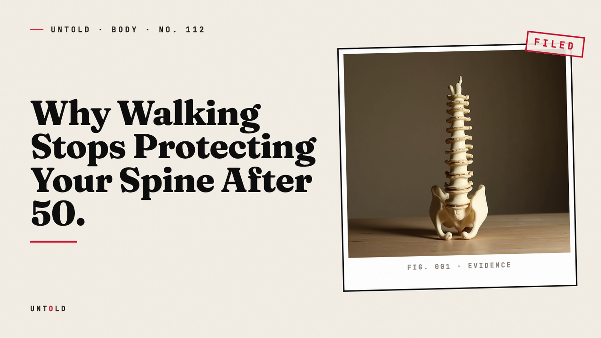
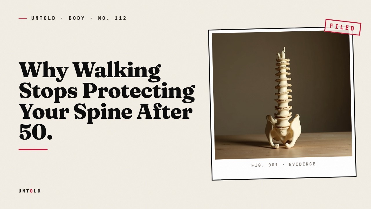
Stand up. Rise onto the balls of your feet, then let your heels drop hard onto the floor. Feel that jolt travel up your shins and into your hips. However small, that shock is one of the few messages your skeleton actually registers. A gentle walk around the block, the exercise most of us have been told keeps our bones strong, does not deliver anything like it.
This is an uncomfortable idea, because walking has been sold to the aging public as the safe, sensible answer to almost everything. Cardiologists prescribe it. Endocrinologists recommend it for blood sugar. Psychologists point to its effect on mood. All of that is real and worth keeping. But somewhere in the diffusion of good advice, walking also acquired a reputation it does not deserve: as weight-bearing exercise capable of holding off the slow thinning of bone that begins in midlife. For the vertebrae stacked along your spine, that reputation is close to fiction.
The consequences are not abstract. The most common fracture of aging is not the hip, as many assume, but the spine. Vertebral compression fractures, in which a weakened bone in the spinal column collapses under ordinary load, affect an estimated seven hundred thousand people a year in the United States alone 1. Many happen without a fall, without drama, sometimes without the person even noticing at first. A vertebra simply gives way, height is lost, posture curves forward, and the quiet arithmetic of frailty advances. The strange part is that the people this happens to are often doing exactly what they were told. They are walking.
The Sensor Buried in Your Skeleton
To understand why walking fails the spine, you have to abandon the idea that bone is inert. We tend to picture the skeleton as a dry scaffold, a set of structural beams that hold up the soft machinery of the body and otherwise do nothing. In truth, bone is among the most metabolically busy tissues we have. It is torn down and rebuilt continuously, throughout life, in a process called remodeling. Specialized cells called osteoclasts dissolve old bone, and osteoblasts lay down new material to replace it. In a healthy young adult, these two processes stay in rough balance. After midlife, and especially after menopause in women, the balance tips toward loss.
What decides where and whether new bone gets built? The answer sits deep inside the mineral itself, in a population of cells most people have never heard of. When an osteoblast finishes laying down bone, some of them become entombed in the very matrix they created. Buried alive, they transform into osteocytes: star-shaped cells with long, fine projections that reach through tiny channels in the bone to touch their neighbors. Threaded together, they form a vast communication network. There are roughly forty billion of them woven through the adult skeleton, and for a long time nobody understood what they were for.
We now know they are sensors. Osteocytes are the strain gauges of the skeleton, the cells that feel mechanical load and translate it into biological instruction. When a force bends a bone, even by an amount far too small to see, fluid shifts through the microscopic channels around these cells. That movement of fluid deforms the osteocyte, and the cell responds by releasing signaling molecules. One of the most important is a protein called sclerostin, which normally acts as a brake on bone formation. When an osteocyte senses meaningful strain, it dials down sclerostin, and the brake comes off. Bone-building cells go to work. In effect, the osteocyte tells the body: something is loading this bone, make it stronger here 2.
There is a crucial catch, and it is the hinge on which this entire story turns. The osteocyte does not respond to load in general. It responds to load that exceeds what the bone is already accustomed to. Strain that falls within the skeleton’s habitual range registers as background noise. The cell has, in a sense, already accounted for it. To provoke a signal, the load has to rise above the customary level, to represent something the bone has not recently felt. Routine, familiar force produces almost nothing.
A Law Written in the Nineteenth Century
The intuition behind this is more than a century old, though the mechanism took most of that century to pin down. In Berlin in the 1890s, a German surgeon named Julius Wolff was studying bones that had been deformed by injury and disease. Examining their internal structure, he noticed something striking. The delicate lattice of trabecular bone inside a joint was not random. Its struts and plates aligned with the lines of mechanical stress the bone habitually carried, as if the tissue had arranged itself to meet the forces demanded of it. Wolff formalized this observation into what became known as Wolff’s Law: bone adapts its architecture to the loads placed upon it 3.
Wolff’s Law described what bone did, but it could not say how. It implied some kind of internal sensor, a way for the tissue to know what forces it was under and respond accordingly, yet the identity of that sensor stayed hypothetical for nearly a hundred years. The person who did more than anyone to close the gap was a British physiologist named Lance Lanyon. Across several decades of careful experiment, Lanyon and his colleagues measured exactly what kinds of load caused bone to grow and what kinds left it unmoved. Their findings overturned a comfortable assumption.
Bone, Lanyon showed, does not respond to slow, repetitive, familiar loading. It responds to loads that are dynamic, high in magnitude, and, above all, unusual. A load applied quickly matters more than the same load applied gradually. A load that arrives in a novel direction or at a novel intensity matters more than one the bone endures every day. The signal for growth fires only when strain crosses above the skeleton’s habitual threshold 4. Lanyon’s work gave the osteocyte its job description before the cell biology had fully caught up, and it explained, in a single stroke, why the exercise most people trust to protect their bones does so little.
Consider what walking actually asks of your legs and spine. Your skeleton already carries your body weight through every waking hour. Standing, shifting, climbing stairs, crossing a room: these are the loads your bones live inside constantly. A walk, however brisk, is more of the same. The forces involved sit comfortably within the range your osteocytes have long since normalized. To those forty billion sensors, a familiar daily walk is not a message. It is silence.
What the Walking Trials Actually Found
This is not merely a theoretical prediction. It has been tested repeatedly, and the results are consistent enough to be disappointing. In 2008, a researcher named Sarah Martyn-St James pulled together the available evidence in a systematic review, gathering the controlled trials that had asked postmenopausal women to walk in order to protect their bones. This is exactly the population most at risk of vertebral fracture, and walking is exactly the intervention they are most often prescribed.
When the studies were pooled, walking produced no significant benefit for the lumbar spine 5. There were hints of a modest effect at the hip, where the geometry and loading of gait may occasionally cross into useful territory. But the spine, the site of the most common fracture of aging, showed nothing meaningful. The gentle, rhythmic, endlessly repeated motion of walking simply never rose above the osteocyte’s threshold in the vertebrae. The bone that most needed defending received no instruction to defend itself.
It is worth sitting with how counterintuitive this is against the public health messaging of the last forty years. We have been told to walk for our bones with the same confidence we have been told to walk for our hearts. For the heart, the advice is sound. For the vertebral spine, the evidence says the effort, however admirable, does not reach the tissue it is aimed at. The spine needed a bigger jolt than any walk could deliver, and no amount of walking was going to supply it.
So researchers began to ask an obviously provocative question. If gentle, habitual load does nothing, what happens if you go to the opposite extreme? What if, instead of protecting fragile bones by keeping loads small, you deliberately made the loads large?
The Trial Nobody Was Supposed to Run
The conventional answer to that question was a flat refusal. Heavy resistance training, the kind that involves loading a barbell to near-maximal weights, was considered reckless for older women with thinning bones. The fear was intuitive. If a vertebra can collapse under ordinary load, surely placing a heavy weight across the shoulders is asking for catastrophe. For years, that fear kept the experiment from being run.
In Queensland, Australia, a bone researcher named Belinda Beck decided to run it anyway. Working with a colleague, Steven Watson, at Griffith University, Beck designed a study that many of her peers considered too dangerous to attempt. She recruited 101 postmenopausal women with low bone mass, women whose average age hovered around sixty-five and whose scans placed them squarely in the population that clinical guidelines warned to avoid heavy lifting altogether. Then she put barbells in their hands 6.
The program was called LIFTMOR, short for Lifting Intervention For Training Muscle and Osteoporosis Rehabilitation, and it did not compromise. Twice a week, under close supervision, the women performed the deadlift, the back squat, and the overhead press, the three heaviest compound lifts in the training vocabulary. They did not lift light. The protocol had them working above eighty-five percent of their one-repetition maximum, which is to say near the upper limit of what their muscles and bones could produce. These were high-magnitude, dynamic, thoroughly unfamiliar loads, precisely the kind of stimulus Lanyon’s work predicted would wake the osteocyte. The trial ran for eight months. A comparison group did a low-intensity home program of gentle movements instead, the sort of exercise usually recommended for women their age.
When the eight months were up and the bone scans came back, the contrast was stark. The women who had lifted heavy gained 2.9 percent in lumbar spine bone mineral density. Over the identical stretch of time, the gentle-exercise group lost 1.2 percent 6. One protocol built the spine. The other could not even hold the line against the ordinary attrition of age. The women lifting near their maximum were doing exactly what everyone had feared would break them, and their spines were getting denser for it.
The Fear That Held Everyone Back
The obvious objection to LIFTMOR is safety, and it deserves a straight answer. Surely, in a group of older women with low bone mass lifting to near-maximal loads, someone got hurt. Across the entire trial, the tally of adverse events attributable to the heavy program amounted to one episode of minor back spasm and a couple of other minor complaints 6. There were no fractures. The catastrophe that had kept the experiment off the table for decades simply did not materialize, provided the lifting was properly taught, properly supervised, and properly progressed.
This reframes the whole picture. The received wisdom had it backward. Gentle motion was thought to be protective and heavy load dangerous. For the vertebral spine, gentle motion turned out to be functionally inert, and heavy load turned out to be the thing that actually built bone, with a safety record that held up under scrutiny. The osteocyte does not care about our caution. It answers only to force that surprises it, and it will keep the skeleton weak until it gets some.
None of this means walking is worthless. It remains one of the finest things a person can do for the cardiovascular system, for metabolic health, for mood and cognition and the simple pleasure of moving through the world. The point is narrower and more precise. Walking is superb for many organs and nearly silent to the vertebral spine. If you want to defend the bones of your back, you have to give them something they do not already expect: loads that are heavy, or fast, or novel enough to cross the threshold. That is why lifting works, and why jumping and hopping, which slam high forces through the skeleton in an instant, can speak the same language a walk never will.
Bone, in the end, is a conversation, and a demanding one. It listens only when you raise your voice. There is a real caution attached to all of this, and it should not be skipped: anyone with diagnosed osteoporosis should never begin heavy loading without a qualified professional guiding the form and the progression, because the margin for poor technique is smaller in a fragile skeleton. But the deeper lesson stands. The next time you rise onto your toes and drop your heels hard against the floor, something genuinely happens beneath the surface. Forty billion sensors register a jolt they were not braced for, and somewhere in the quiet architecture of your spine, cells decide to build.
Sources
- Burge, R. et al., Incidence and Economic Burden of Osteoporosis-Related Fractures, Journal of Bone and Mineral Research, 2007. — https://asbmr.onlinelibrary.wiley.com/doi/10.1359/jbmr.061113
- Bonewald, L. F., The Amazing Osteocyte, Journal of Bone and Mineral Research, 2011. — https://asbmr.onlinelibrary.wiley.com/doi/10.1002/jbmr.320
- Wolff, J., Das Gesetz der Transformation der Knochen (The Law of Bone Remodeling), 1892. — https://en.wikipedia.org/wiki/Wolff%27s_law
- Lanyon, L. E. & Rubin, C. T., Static vs Dynamic Loads as an Influence on Bone Remodelling, Journal of Biomechanics, 1984. — https://www.sciencedirect.com/science/article/abs/pii/0021929084900035
- Martyn-St James, M. & Carroll, S., A meta-analysis of impact exercise on postmenopausal bone loss: the case for mixed loading exercise programmes, British Journal of Sports Medicine, 2009. — https://bjsm.bmj.com/content/43/12/898
- Watson, S. L., Weeks, B. K., Beck, B. R. et al., High-Intensity Resistance and Impact Training Improves Bone Mineral Density and Physical Function in Postmenopausal Women With Osteopenia and Osteoporosis: The LIFTMOR Randomized Controlled Trial, Journal of Bone and Mineral Research, 2018. — https://asbmr.onlinelibrary.wiley.com/doi/10.1002/jbmr.3284
Related reading