The Forest Inside Your Chest
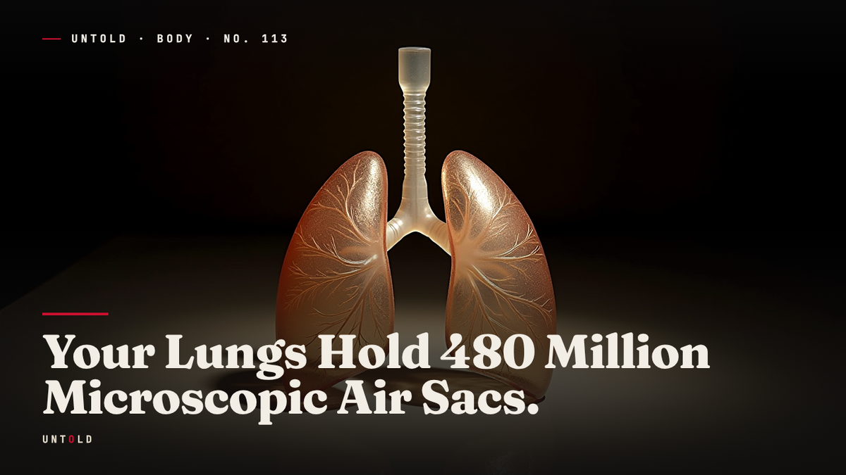
Your lungs are not two pink bags but a foam of 480 million sacs, and they came before land.
Take the deepest breath you can manage. Hold it a moment. The air you just pulled inward is not sitting in a pair of hollow chambers, waiting to be pushed back out. It has been distributed, in an instant, across a surface roughly the size of a small studio apartment: a sprawling, wet, folded landscape hidden entirely inside the cage of your ribs. Every breath floods it. Every breath drains it. And most of us go a lifetime without any picture of what is actually happening in there.
The picture most people carry is of two soft pink bags, balloons that swell and shrink with each breath. It is a serviceable image for a children’s book and almost entirely wrong. A lung is not a bag. It is a foam. It is a dense, branching thicket of millions of microscopic chambers, each one a fraction of a millimeter across, each one wrapped in blood vessels thinner than anything else in the body. If you could unpack that foam and lay it flat, it would not fit on a table. It would cover the floor of a room.
And there is a stranger fact still, one that upends the neat story most of us learned in school. The lung did not evolve so that animals could crawl out of the water and breathe air on land. The lung came first. It was already there, deep in our fish ancestry, long before anything grew legs. The organ in your chest is older than the idea of dry ground.
A Frog, a Lens, and the End of the Bag
To understand the breath, it helps to start in 1661, with an Italian anatomist and one of the first useful microscopes.
Marcello Malpighi, born near Bologna in 1628, belonged to the first generation of scientists who could see below the threshold of the naked eye. The microscope was new, crude by later standards, and its images swam and blurred. But it revealed a world that no dissection knife could reach. Malpighi turned this new instrument on the body’s tissues one after another, and in the process he founded the study of anatomy at the microscopic scale 1.
When he placed a frog’s lung beneath his lens, he expected, as everyone did, to find dense, uniform tissue. Solid meat. Instead he found something that looked almost impossible: a network of tiny hollow chambers, thin-walled, threaded through with the finest blood vessels. The lung was not a solid organ and not a simple bag. It was a mesh of countless small sacs, each embraced by a web of capillaries so fine they had never been described before. Malpighi had, in the same act, discovered both the alveolus and the capillary bed that serves it 2.
This was the true engine of breathing, hiding in plain sight. Not the lung as a whole, not the visible bellows of the chest, but the alveolus: the terminal air sac where the actual work happens. It is here that oxygen slips out of the air and into the blood, and carbon dioxide moves the other way. Everything else in the respiratory system, the windpipe, the branching bronchi, the muscular effort of the diaphragm, exists only to deliver fresh air to these millions of tiny chambers and to carry the stale air away. The foam is the point. The bag is just the container.
The Long Problem of Counting
Malpighi could see the sacs. He could not count them. Nor could anyone else for the next three centuries.
The difficulty is not conceptual but practical, and it is severe. A single human alveolus measures around two-tenths of a millimeter across, thinner than a grain of salt. They are packed together in three dimensions, layered and overlapping, their walls shared between neighbors like the cells of a honeycomb. You cannot simply slice a lung open and tally the holes on the cut surface, because a two-dimensional slice through a three-dimensional foam gives you a systematically distorted count. Some sacs are caught in cross-section, some in glancing tangents, some missed entirely. For a long time, estimates of how many alveoli a human lung contained ranged wildly, from a few million to well over a billion. The honest answer was that nobody knew.
The number resisted for so long because counting objects inside a solid volume is a genuinely hard mathematical problem, and the tempting shortcuts all introduce bias. If you count only the sacs that happen to be big on your slice, you overcount the big ones. If you count profiles rather than particles, you count the same sac twice. A whole subfield of quantitative anatomy grew up around exactly this difficulty, and its answer was a discipline called design-based stereology: a set of statistical sampling methods that let you estimate the number and size of structures in three dimensions without the distortions of naive counting 3.
The principle is elegant. You slice the tissue in a carefully randomized way, you sample small volumes according to unbiased rules, and you scale those samples up using mathematics designed to be immune to the shape and orientation of what you are counting. Done properly, it gives a number you can defend, along with an honest measure of how uncertain that number is. The method removed the guesswork, but it demanded patience of an almost monastic order.
Four Hundred and Eighty Million
Two researchers supplied that patience. Ewald Weibel, an anatomist at the University of Bern, had spent much of his career mapping the hidden architecture of the lung, chamber by chamber and branch by branch. He was, more than anyone, the person who turned the lung into a countable, measurable object rather than a soft mystery. Working with Matthias Ochs and colleagues, he brought the full apparatus of modern stereology to bear on the oldest unanswered question about the organ 4.
In 2004 they published the result. Using design-based stereology on human lungs, they arrived at an average of roughly 480 million alveoli in a single adult pair 4. After three hundred years of guessing, there was finally a number with a method behind it.
But the number came with a caveat that made it more interesting, not less. It was not fixed. Across the lungs they examined, the count ranged from about 274 million to nearly 790 million, a spread of almost threefold from one person to the next 4. The variation was not random noise. Larger lungs held more sacs. The alveolar count tracked closely with total lung volume, which meant that a tall person with a large chest was likely carrying a genuinely different quantity of breathing surface than a small one. There is no single canonical human lung. There is a distribution, and 480 million is simply its middle.
Now perform the imaginative act that the number invites. Take those hundreds of millions of sacs, each a fraction of a millimeter wide, and unfold them. Flatten the entire foam into a single continuous sheet. The surface it covers is around seventy square meters, roughly the floor area of a modest studio apartment 5. That entire expanse is folded, compressed, and tucked into two handfuls of tissue behind your breastbone. It is one of the most extreme feats of packing in the body: nature’s solution to a simple engineering problem, which is to fit the maximum possible surface into the minimum possible space.
Surface is everything, because gas exchange happens across surface. Every one of those sacs is sheathed in the thinnest blood vessels you possess. The barrier between air and blood, the membrane that oxygen must cross to reach your bloodstream, is on the order of a thousandth of a millimeter thick, so delicate that it is almost more a film than a wall 4. In the time it takes your heart to beat once, air and blood are brought face to face across that gossamer film, and oxygen crosses in one direction while carbon dioxide crosses in the other. Twenty thousand times a day, the whole vast surface is refreshed.
The Story That Runs Backwards
That is the architecture. Its origin is where the familiar account collapses.
The story most of us absorbed is clean and satisfying. Fish had gills. Life began in the water. When vertebrates ventured onto land, they needed a new way to breathe air, and the lung was the innovation that made the move possible. Gills for water, lungs for land, and the lung was the ticket ashore. It has the shape of a good story: a problem, an invention, a triumph.
It is backwards.
Comparative anatomists, tracing the lung across the whole family of vertebrates, kept finding it in the wrong place. They found lungs, or organs unmistakably descended from lungs, in bony fish that never left the water at all and never had any ancestor that did. Air sacs branching off the gut, capable of holding gulped air, turned up across ancient lineages of fish. The lung was not a novelty invented at the water’s edge. It was already present, deep in the fish body plan, long before any vertebrate had legs 6.
Why would a fish, surrounded by water, want a lung? Because water is not a reliable place to find oxygen. Warm water holds less dissolved oxygen than cold water, and stagnant pools, swamps, and slow tropical rivers can become nearly anoxic. A fish that can rise to the surface and gulp a mouthful of air into an internal sac has an escape route when the water itself starts to suffocate. Many living fish do exactly this, and the lungfishes, which survive droughts by breathing air in mud burrows, are only the most dramatic example of an ancient and widespread habit. The primitive lung was not a device for leaving water. It was a device for surviving bad water.
Then comes the twist within the twist. The comparative anatomist Karel Liem, working at Harvard, was among those who followed this logic to its conclusion by looking at the swim bladder, the gas-filled organ that lets most modern fish hover weightlessly at a chosen depth 7. The intuitive assumption is that the lung must be a modified swim bladder, a fish organ repurposed for air. The truth is the reverse. The swim bladder is the derived structure, evolved from the ancestral lung. The buoyancy float that lets a fish hang motionless in open water is a repurposed breathing organ, a lung that stopped breathing and learned to provide lift instead 67.
The order of events, then, runs opposite to the schoolbook version. The lung came first, roughly 400 million years ago, in fish that lived and stayed in the water 6. From that ancient air sac, two lineages diverged. In one, the lung remained a lung and was carried, unchanged in its essential design, up onto the land and forward through the whole history of four-limbed animals, down to the foam inside your ribs. In the other, the same organ was co-opted into a buoyancy device, and those fish went on to fill the oceans. The lung is not the thing that got us out of the water. It is the thing we brought with us.
An Inheritance, Not an Invention
Hold all of this together and look again at a single breath.
You are not inflating two pink bags. You are flooding a forest of nearly half a billion microscopic sacs, spreading a thin film of air across a surface the size of a room, pressing it against blood through a membrane a thousandth of a millimeter thick, and doing it again twenty thousand times before the day is out. The elegance of the thing is total, and it is almost entirely hidden.
And the architecture doing this work was not designed for the life you live. It was not built to conquer the land. It was carried out of the water by animals that already had it, animals for whom it was an emergency measure against suffocating pools. The land came later. The legs came later. The lung was already there, an old solution to an old problem, waiting.
So the next time you take a deep breath and feel your chest expand, it may be worth remembering what that sensation actually is. Not the filling of two simple containers, but the refreshing of an immense folded surface, an inheritance from the sea that predates the ground you are standing on. Four hundred and eighty million tiny answers, on average, to a single ancient question: how does a body reach the air.

Sources
- Malpighi, M., De pulmonibus observationes anatomicae, 1661. — https://en.wikipedia.org/wiki/Marcello_Malpighi
- West, J. B., “Marcello Malpighi and the discovery of the pulmonary capillaries and alveoli,” American Journal of Physiology - Lung Cellular and Molecular Physiology, 2013. — https://journals.physiology.org/doi/full/10.1152/ajplung.00016.2013
- Weibel, E. R., “Stereological Methods,” Academic Press, 1979. — https://en.wikipedia.org/wiki/Stereology
- Ochs, M., Weibel, E. R. et al., “The number of alveoli in the human lung,” American Journal of Respiratory and Critical Care Medicine, 2004. — https://www.atsjournals.org/doi/full/10.1164/rccm.200308-1107OC
- Weibel, E. R., “Morphometry of the Human Lung,” Springer, 1963. — https://link.springer.com/book/10.1007/978-3-642-87553-3
- Longo, S. J. et al., “Homology of lungs and gas bladders: insights from arterial vasculature,” Journal of Morphology, 2013. — https://onlinelibrary.wiley.com/doi/10.1002/jmor.20146
- Liem, K. F., “Form and function of lungs: the evolution of air breathing mechanisms,” American Zoologist, 1988. — https://academic.oup.com/icb/article/28/2/739/181495
Related reading