The Calendar Written in Blood Beneath Your Skin
A bruise is not damage on display. It is the visible record of your body dismantling and recycling itself.
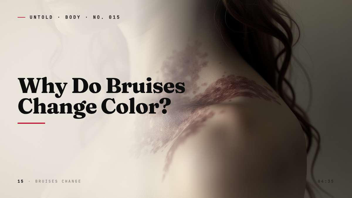
A coffee table is a quiet enemy. You walk past it a thousand times without incident, and then one evening your shin finds its corner, and for a moment the world narrows to a single point of pain. Hours later, something appears on the skin: a faint pinkish smudge that you might not have noticed if you weren’t looking. By morning it has deepened into a bruise the color of a thundercloud. And then, over the following days, it begins to do something genuinely strange. The purple drains away into blue. The blue softens into a sickly green. The green yellows like an old photograph. And by the end of the second week, the mark has faded into nothing, leaving the skin exactly as it was before.
We tend to regard bruises as passive things, as injuries we wait out. But that procession of colors is not the slow decay of a wound. It is the opposite. Each shade marks a distinct chemical reaction unfolding beneath the surface, a sequence so reliable that forensic pathologists once believed they could read a bruise like a clock and tell you, to the day, when a person had been struck. The rainbow on your arm is a record of labor. It is your body taking apart spilled blood, molecule by molecule, and carrying the pieces away.
A Flood Beneath Intact Skin
The story begins with rupture. When something strikes the body hard enough, the force is transmitted through the skin into the soft tissue below, and there it tears open the smallest blood vessels in the body: the capillaries. These are vessels of almost unimaginable delicacy. The thinnest of them are narrower than a single red blood cell, and their walls are just one cell thick, a single layer of endothelium with nothing to reinforce it 1. They are built for the gentle business of exchange, handing oxygen to tissue and collecting carbon dioxide in return, not for absorbing the blow of a hardwood edge.
So they break. Blood escapes from the broken vessels and seeps into the surrounding tissue, but because the skin itself remains unbroken, none of it can leave the body. It simply pools, trapped in the spaces between cells. Doctors call this collection of escaped blood a hematoma when it is large enough, and a contusion, or simply a bruise, when it is the more familiar diffuse stain. The blood is now in the wrong place. It is no longer circulating, no longer carrying oxygen anywhere useful. It is, from the body’s point of view, debris.
For the first few hours, the bruise looks pink or red, sometimes barely visible. That color is fresh blood, still oxygenated, sitting close enough to the surface to show its true hue through the skin. But oxygen is a perishable cargo. The hemoglobin inside red blood cells carries it bound to iron, and once those cells are stranded outside the circulation, there is no fresh supply coming. The oxygen is consumed or lost, and the hemoglobin that remains is now deoxyhemoglobin, a darker, bluer pigment. This is the same molecule responsible for the bluish appearance of the veins on the back of your hand. Seen through layers of skin and tissue, deoxygenated blood does not look red at all. It looks deep purple, almost black. This is the bruise at its most dramatic, the stage we think of as a proper bruise, and it signals that the body now faces a cleanup operation of considerable precision.
The First Yellow Clue
The science of what happens next begins, oddly enough, in the mid-nineteenth century, with one of the founders of modern medicine peering at old blood. Rudolf Virchow, the German pathologist who would later coin the principle that every cell arises from another cell, was studying the residue left behind by hemorrhages. In 1847 he identified a yellow crystalline pigment in old clots and bruised tissue, a substance he named hematoidin 2. It was, in effect, the first chemical signature of a healing wound, physical proof that the body was not merely waiting for trapped blood to disappear but was actively transforming it into something new.
Virchow’s insight was that the bruise was a site of work. The blood was being dismantled. What he could not yet see, because the tools did not exist, was the machinery doing the dismantling. That machinery turned out to be cellular, and it arrives at the scene like a demolition crew.
Within a day or two of the injury, the immune system dispatches its scavengers. The most important of these are the macrophages, a class of white blood cell whose name comes from the Greek for “big eaters,” and the name is literal. A macrophage is a roving cell that engulfs and digests whatever the body has flagged as waste: dead cells, microbes, foreign particles, and, in the case of a bruise, the stranded red blood cells leaking from the broken capillaries 3. The macrophage wraps itself around the damaged cell, swallows it whole into an internal compartment, and begins to break it apart from the inside.
This is where the colors come from. Each shade of a healing bruise corresponds to a stage in the chemical breakdown of hemoglobin, the very molecule that gave the fresh blood its red.
A Rainbow Made of Chemistry
Inside the macrophage, hemoglobin is taken apart in an orderly sequence, and the body extracts a series of compounds from it, each with its own color. The first transformation produces biliverdin, a green pigment. Biliverdin is what gives a bruise that faintly nauseating greenish tinge that tends to appear around the fourth to seventh day, when the worst of the purple has begun to lift 4. If you have ever looked at an aging bruise and noticed a band of green at its edges, you were watching this reaction in progress. It is the same green that appears in the skins of certain bruised fruits and, for the same biochemical reason, in the bile of many animals.
Biliverdin does not last. An enzyme converts it into bilirubin, a yellow-brown pigment, and this is the source of the yellow stage that follows the green. Bilirubin is a molecule with a wider fame than most people realize. It is the same compound that accumulates in newborns whose livers have not yet learned to process it efficiently, giving their skin the yellow cast of jaundice. When you watch a bruise turn yellow, your tissue is manufacturing the identical chemical, in miniature, and then carrying it away to be excreted. The bruise, in a real sense, is a wound healing in slow motion, and the yellow is the color of that healing reaching its final phase.
There is one more pigment in the sequence, and it represents the part of the story that is easiest to overlook. Hemoglobin is built around iron, four atoms of it per molecule, and iron is too valuable for the body to throw away. As the macrophages finish their work, the iron is stored in a golden-brown compound called hemosiderin. This is not waste in transit. It is salvage. The iron held in hemosiderin can be returned to the body’s reserves and used again, in new red blood cells, in new hemoglobin, to carry oxygen on some future day 5. The faint brownish stain that lingers at the end of a bruise’s life is the body refusing to let anything go to waste, recycling the raw material of one broken cell into the machinery of the next.
Laid out in order, the sequence reads almost like a poem of decomposition: red, then purple and blue, then green, then yellow, then brown, then nothing. The average bruise completes this journey in something like ten to fourteen days, a procession so consistent that it can feel less like injury than like a process running to schedule 6. Which is exactly what led a generation of medical examiners to a tempting and ultimately troublesome conclusion.
Reading Bruises Like Clocks
If the colors of a bruise unfold in a fixed order over a predictable span of days, then a bruise should be readable in reverse. Show a pathologist a yellow-green mark and they ought to be able to say it was inflicted roughly a week ago. Show them a fresh purple one and they should date it to the past day or two. This logic mattered enormously in the courtroom. In cases of homicide, assault, and child abuse, the age of an injury can corroborate or destroy an alibi, can place a suspect at a scene or rule them out, can distinguish a single violent episode from a sustained pattern of harm.
Forensic pathology took this task seriously. Vincent Di Maio, an American medical examiner whose textbooks on gunshot wounds and forensic pathology trained generations of investigators, examined countless bruises over a long career and helped establish the conventions by which injuries were dated by color 7. For decades, the color chart of the healing bruise functioned in forensic practice as something close to a timestamp, a way of converting the appearance of skin into a number of days.
The trouble was that the chart had never been properly tested. It was an accumulation of clinical impression and received wisdom, repeated from textbook to textbook, rather than a finding built on controlled observation. And when someone finally did test it, the edifice came apart.
When the Calendar Broke
In 1996, the British forensic scientist Peter Vanezis, working at the University of Glasgow, reviewed the existing literature on dating bruises by their color, and what he found was not a calendar but chaos 8. Later, rigorous studies confirmed the problem with uncomfortable clarity. In one frequently cited investigation, examiners were asked to judge the age of bruises from photographs and from direct observation, and their estimates were so inconsistent, both from one examiner to another and against the known true age of the injuries, that the practice could not be defended. A separate landmark study established the single most damaging finding of all: a yellow color, long treated as the reliable marker of an older bruise, could appear in some bruises within eighteen hours and might never appear at all in others 9.
The reason is that the timeline is not fixed at all. It is exquisitely sensitive to the individual body. The same blow, delivered to two different people, can produce bruises that pass through their colors at entirely different speeds. The depth of the injury matters: a bruise deep in the tissue may not become visible at the surface until days after the impact, by which point its internal chemistry has already advanced. Skin tone matters, changing how each pigment reads through the surface. Age matters enormously. And the color a bruise presents depends on the lighting, the photograph, and the eye of the observer.
So the confident science of dating bruises by color was quietly retired, or at least heavily qualified. What survived was the underlying biochemistry, which never depended on the timing being uniform. The pigments still appear in the same order in every body, because they are the products of the same chemical pathway. It is only the clock that varies, not the chemistry.
Why No Two Bruises Are Alike
The individuality of bruising becomes most obvious at the extremes of age. Older skin bruises far more easily than young skin, and it bruises from contact so minor it might pass unnoticed in a younger person. The reasons are structural. With age, the capillaries become more fragile and the supportive scaffolding around them thins out. Collagen, the protein that gives skin its resilience and cushions the small vessels against impact, diminishes over the years, leaving those one-cell-thick capillary walls with less protection from the forces of ordinary life. The fat layer beneath the skin, which acts as padding, also grows thinner. The result is that a bump which would leave a younger person unmarked can open the capillaries in an older one. Among adults over sixty-five, a substantial fraction bruise after only minor contact, and the resulting marks often take longer to clear 10.
The timeline can be stretched by other factors too. Blood-thinning medications, by their very design, make it harder for the body to seal off the leaking vessels, so more blood escapes and the bruise is larger and slower to fade. Deficiencies in vitamins involved in clotting and in the maintenance of vessel walls can prolong the rainbow. So can a range of underlying conditions affecting the blood or the connective tissue. None of these change the colors themselves. They change only the pace at which the colors arrive and depart, which is precisely why a single universal calendar was never going to work.
What remains constant is the meaning of the sequence. A bruise, properly understood, is not a wound on display. It is a project: a rupture, a flood, the arrival of a cleanup crew, the patient disassembly of hemoglobin into biliverdin and then bilirubin, the salvage of iron into hemosiderin, and finally the restoration of the tissue to its original state, with the reclaimed iron returned to circulation and the spent pigments excreted from the body. Nothing of the spilled blood is left behind. Nothing is wasted.
There is something worth pausing over in that. The body conducts this entire operation without instruction and almost entirely out of sight. We notice only the byproduct, the changing color that happens to fall within the narrow band of light our eyes can read, while the actual work proceeds in the dark of the tissue. Healing is, for the most part, invisible. The bruise is one of the rare occasions when it is briefly made visible, when the repair leaves a stain we can watch.
So the next time a purple mark blooms on a shin or a forearm, it is worth looking closely, and looking again over the following days. The thundercloud will lift into blue, the blue will turn green, the green will yellow, and the yellow will fade into clean skin. You will not be watching a wound. You will be watching your own body take blood apart, sort it into its component pigments, recover what it can use, and rebuild the tissue around the wreckage. It is a calendar written in iron and light, and it keeps perfect chemical order even when it refuses to keep time.

Sources
- Tortora, G. J., and Derrickson, B., Principles of Anatomy and Physiology (capillary structure), Wiley, 2017. — https://www.wiley.com/en-us/Principles+of+Anatomy+and+Physiology%2C+15th+Edition-p-9781119343738
- Virchow, R., on hematoidin and the breakdown of extravasated blood, Archiv fur pathologische Anatomie, 1847. — https://www.ncbi.nlm.nih.gov/pmc/articles/PMC4322417/
- Gordon, S., and Pluddemann, A., “Macrophage clearance of apoptotic cells and red blood cells,” Frontiers in Immunology, 2018. — https://www.frontiersin.org/articles/10.3389/fimmu.2018.02012/full
- Stevenson, T., “The colour changes of bruises and their interpretation,” review of hemoglobin breakdown pigments, Journal of Clinical Forensic Medicine, 2002. — https://pubmed.ncbi.nlm.nih.gov/15275006/
- Ganz, T., “Macrophages and iron metabolism,” Microbiology Spectrum, 2016. — https://journals.asm.org/doi/10.1128/microbiolspec.MCHD-0037-2016
- Hughes, V. K., et al., “The practical application of reflectance spectrophotometry for the demonstration of haemoglobin and its degradation in bruises,” Journal of Clinical Pathology, 2004. — https://jcp.bmj.com/content/57/4/355
- Di Maio, V. J. M., and Di Maio, D., Forensic Pathology, 2nd Edition, CRC Press, 2001. — https://www.routledge.com/Forensic-Pathology/DiMaio-DiMaio/p/book/9780849300721
- Vanezis, P., “Interpreting bruises at necropsy,” Journal of Clinical Pathology, 2001 (building on 1996 review of bruise-dating evidence). — https://jcp.bmj.com/content/54/5/348
- Langlois, N. E. I., and Gresham, G. A., “The ageing of bruises: a review and study of the colour changes with time,” Forensic Science International, 1991. — https://pubmed.ncbi.nlm.nih.gov/1748277/
- Mosqueda, L., et al., “The life cycle of bruises in older adults,” Journal of the American Geriatrics Society, 2005. — https://pubmed.ncbi.nlm.nih.gov/16078959/
Related reading