The Smallest Muscle's Confession
What a fluttering eyelid reveals about an overworked nervous system, and when the twitch becomes a warning.
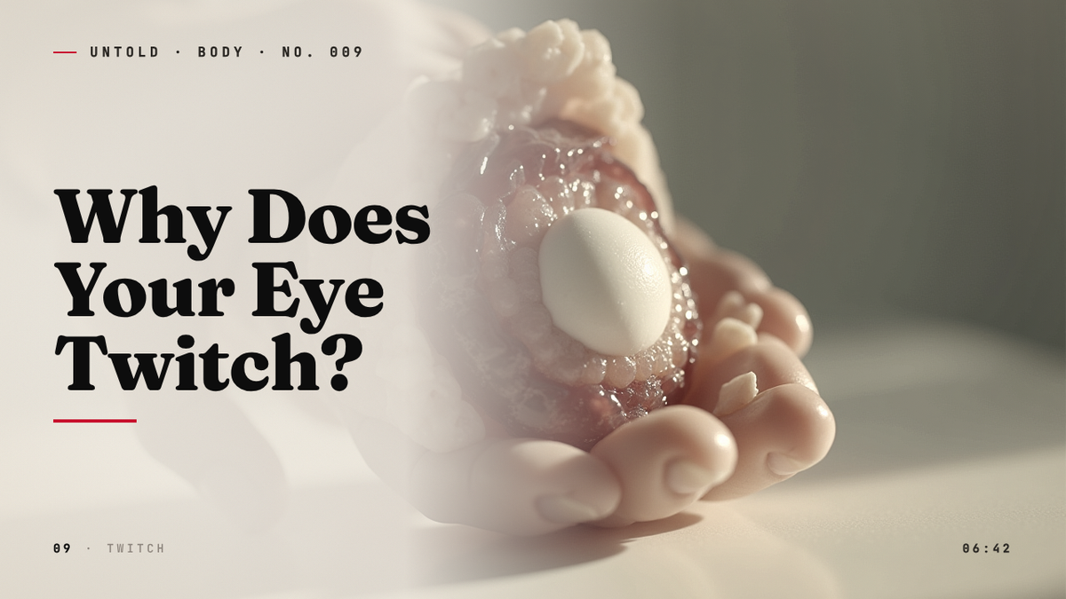
It begins, almost always, in the middle of something else. A meeting. A long drive. The fourth hour of a screen. A flutter starts under one eye, soft and insistent, like a moth trapped behind the skin. You raise a hand to the spot. You glance at the bathroom mirror. Nothing moves. Nothing visible, anyway. But you can feel it, a tap, a tap, a tap, and once you notice it, you cannot un-notice it.
It may last an hour. It may last three days. In stubborn cases, three weeks. For most people who experience it, and surveys suggest the great majority of adults will in any given year, the twitch passes the way a sneeze passes: an irritation, then a memory 1. But the question of what causes it, of why a single sliver of muscle around the eye should suddenly take on a life of its own, opens a small window into the way the human nervous system manages, and occasionally mismanages, its own electricity.
Doctors call it myokymia. From the Greek myo, muscle, and kyma, wave. A small wave of muscle, breaking on its own.
The most active muscle you own
The ring of tissue responsible for closing your eye is called the orbicularis oculi. It encircles each socket like a drawstring, thin as parchment in places, and it is among the most relentlessly used skeletal muscles in the body. A healthy adult blinks somewhere between 14,000 and 19,000 times a day, mostly without noticing 2. Each blink is a small, choreographed electrical event: a signal departs the brainstem, travels along the facial nerve (cranial nerve VII), and arrives at a bundle of muscle fibers that contract in unison, sweeping the eyelid down and back up in roughly a third of a second.
Multiply that by a waking lifetime and the orbicularis oculi performs more discrete contractions than the heart. Unlike the heart, it does so under the watchful, if usually unconscious, supervision of the cortex. Blinking is reflexive, but it is also modifiable. You can suppress a blink during a staring contest. You can exaggerate one in a wink. The muscle answers to the brain.
Which is why, when it begins to answer to no one, it feels uncanny.
In myokymia, a single motor unit, one nerve fiber and the small cluster of muscle fibers it innervates, begins to discharge spontaneously. The cortex has not sent a command. The reflex arc has not been tripped. A fiber simply fires, then fires again, then settles into a rhythm of its own choosing. Because the orbicularis oculi is thin and the eyelid is thinner still, even one rogue fiber produces a sensation entirely out of proportion to its size. It feels enormous. It is, in fact, microscopic. This is part of why the twitch is usually invisible in the mirror: there is barely anything there to see.
A neurologist in the wake of war
The formal description of myokymia traces back to the early twentieth century. The German neurologist Friedrich Schultze had noted continuous muscle rippling under the skin in the 1890s, but the eyelid variant was characterized more fully in the years after the First World War, when European clinics filled with soldiers whose bodies refused to settle 3. Among the complaints were tremors, fasciculations, and persistent eyelid twitches that did not resolve with rest. The men were exhausted in a way that medicine did not yet have good language for. Their nerves, clinicians reasoned, had been left in a state of low-grade overexcitation, as if the dial that turned motor neurons up and down had been bent slightly out of true.
It was a useful intuition, and a durable one. A century later, the dominant explanation for benign eyelid myokymia is still, in essence, an overexcitation story. Something has lowered the threshold at which a motor neuron in the facial nerve fires. The neuron, sitting now closer to its trigger point than usual, occasionally discharges without instruction. The patient does not have a disease. The patient has a nervous system that is running slightly too hot.
The interesting question is: what turned up the dial?
The modern life cocktail
When researchers survey patients who present with persistent eyelid twitching, the same handful of factors appears with such regularity that ophthalmology textbooks list them almost as a refrain: stress, fatigue, caffeine, alcohol, dry eyes, and, more arguably, mineral imbalance 4. None of these are exotic. All of them are, in the modern world, ambient.
Stress is the variable patients most often identify themselves. In clinical surveys of myokymia, a substantial majority of those affected report that the twitching coincided with periods of unusual psychological pressure, and that it subsided within days of the pressure easing. The mechanism is not mysterious. Sustained stress raises circulating levels of cortisol and catecholamines, hormones that, among many other effects, increase the excitability of motor neurons. Muscles do not become stronger under stress; they become twitchier. The smallest muscles, with the fewest fibers and the thinnest margin between rest and recruitment, register the change first. The orbicularis oculi is among the smallest skeletal muscles in the human body. It is, in a sense, the canary in the coal mine of the motor system.
Caffeine works along a parallel pathway. Adenosine is the molecule that accumulates in the brain across a waking day and signals fatigue; one of its quieter functions is to dampen the firing of certain neurons. Caffeine blocks adenosine receptors. The result is alertness, which is the point, but also a generalized loss of inhibition in circuits that depend on adenosine’s calming touch. Heavy coffee drinkers, particularly those who have layered three or four cups on top of poor sleep, report eyelid twitching at notably higher rates than light drinkers 5. Cutting back, in clinical practice, is one of the first interventions a doctor will recommend, and one of the most reliably effective.
Sleep deprivation compounds both effects. The brain’s ability to suppress stray, unwanted motor signals depends on the integrity of inhibitory networks that are themselves restored during sleep. A poorly rested cortex is a leaky cortex. Signals that would normally be filtered out, including the meaningless chatter of an over-caffeinated motor neuron, get through. The eyelid registers the noise.
A twitching eye, in other words, is rarely an isolated problem. It is a small, peripheral readout of a larger condition: a body operating at the edge of its tolerance for stimulation.
Magnesium and the chemistry of relaxation
There is one further variable that deserves a closer look, because it is both unfashionable and unusually plausible. Magnesium.
Muscle contraction is, at its core, a calcium event. When a nerve signal arrives at a muscle fiber, calcium ions flood into the cell and allow the contractile proteins, actin and myosin, to slide against one another. Relaxation requires the calcium to be pumped back out. Magnesium is the ion that competes with calcium at many of the relevant binding sites, and it is essential to the smooth, regulated cycling between contraction and rest. When magnesium runs low, calcium has freer rein. Muscle fibers contract more easily and relax more reluctantly. Spasms, cramps, and fine twitches become more likely.
The American physician and researcher Mildred Seelig spent much of her career arguing that subclinical magnesium deficiency was both more common and more consequential than mainstream medicine acknowledged. In a series of papers and her 1980 monograph Magnesium Deficiency in the Pathogenesis of Disease, she connected low magnesium status to a range of neuromuscular irritabilities, including persistent fasciculations 6. Her broader claims about cardiovascular disease were controversial, and remain debated. But the narrower link to muscle excitability has held up reasonably well, and dietary surveys consistently find that a sizable fraction of adults in industrialized countries fall short of the recommended daily intake.
This does not mean every twitching eyelid is a magnesium problem, or that supplementation is a panacea. Most cases of myokymia resolve before any nutritional intervention has time to register. But for a patient whose twitch has lingered for weeks, whose diet is heavy on processed food and light on leafy greens, and whose other investigations have come back unremarkable, a quiet attention to magnesium intake is a sensible step.
The eye that cannot lubricate itself
There is another category of cause, less hormonal, more mechanical, that has grown more important in the era of screens. The cornea is one of the most densely innervated surfaces in the body. When it dries out, even slightly, those nerves become irritable. The brain registers the irritation as a need to blink, and the orbicularis oculi obliges. If the dryness persists, the system can begin to overshoot, producing a low-grade tic at the eyelid that has nothing to do with stress or caffeine and everything to do with the simple fact that the eye is not being adequately bathed.
The physiology of blinking changes dramatically in front of a screen. Studies dating back to the 1990s found that blink rate drops by as much as half during sustained visual concentration, particularly when reading on a monitor 7. Each blink also tends to be incomplete, the eyelid not quite reaching the lower lid before retracting. The tear film thins. The surface dries. The nerves complain. And in a small percentage of cases, the complaint takes the form of a flutter that arrives, mysteriously, hours after the screen has been closed.
This is myokymia as a kind of protective reflex gone slightly out of tune. The system is trying to help. It is helping a little too eagerly.
When the twitch becomes something else
For the great majority of people, the story ends here. Sleep, water, fewer espressos, a long walk away from the inbox, and within a week or two the eyelid settles. The twitch is, in the technical language of the field, benign. It does not progress. It does not herald disease. It is the nervous system clearing its throat.
There are, however, two conditions that look superficially similar and are not benign, and they are worth understanding clearly, because the distinction is what makes the difference between a minor annoyance and a problem that wants medical attention.
The first is benign essential blepharospasm. The name is misleading: the word benign refers to the fact that the condition is not life-threatening, not to the experience of having it, which can be debilitating. In blepharospasm, both eyelids contract involuntarily and forcefully, sometimes squeezing shut for seconds at a time, sometimes for long enough that the person is functionally blind during the episode. It typically begins in middle age, affects women more often than men, and is estimated to involve on the order of fifty thousand people in the United States 8. Unlike garden-variety myokymia, it does not respond to a good night’s sleep. The current best treatment is targeted injection of botulinum toxin into the offending muscles, which weakens them enough to prevent the squeeze without paralyzing the eye.
The second is hemifacial spasm. Here the twitching is unilateral, but it does not stay confined to the eyelid. Over months or years it spreads downward, recruiting the cheek, then the corner of the mouth, eventually the platysma of the neck. The cause, in most cases, is anatomical: a small artery or vein at the base of the brain has come to lie against the facial nerve where it exits the brainstem, and each pulse of blood through that vessel irritates the nerve. The irritation, repeated millions of times, eventually produces a pattern of spontaneous discharge that no medication can fully suppress.
The definitive treatment for hemifacial spasm was developed by the American neurosurgeon Peter Jannetta, who in 1966 began performing what he called microvascular decompression: a delicate operation in which the offending vessel is identified, lifted gently away from the facial nerve, and held in its new position by a small pad of inert material 9. The results, in skilled hands, were and remain remarkable. Patients whose faces had spasmed for decades woke from surgery still. Jannetta’s procedure took years to be accepted by a skeptical neurology establishment, in part because the idea that a great deal of human misery could be caused by a single misplaced blood vessel struck many of his contemporaries as too simple to be true. It turned out to be true.
These conditions are rare. The point of mentioning them is not to alarm the reader whose eyelid has been fluttering for two days. It is to mark, clearly, the line at which a twitch crosses into something a clinician should see.
When to actually worry
Among neurologists and ophthalmologists, a rough consensus has emerged about what features of an eyelid twitch deserve closer attention. The list is short and worth keeping in mind.
A twitch that persists for more than three weeks without easing. A twitch that spreads beyond the eyelid to involve the cheek, the mouth, or the neck. A twitch accompanied by drooping of the eyelid, partial closure of the eye, double vision, blurring, weakness elsewhere in the face, or any change in sensation. Any of these warrants a visit to a neurologist or an ophthalmologist, not because catastrophe is likely, but because the differential diagnosis at that point becomes wide enough that it is worth a professional’s attention.
For everyone else, which is to say almost everyone, the twitch is a message rather than a diagnosis. It is the nervous system, in its small and stubborn way, registering a complaint that the conscious mind has been too busy to file. Most people, asked directly, can name what the twitch is about. They have been sleeping six hours a night for two weeks. They have been on their third coffee by ten in the morning. They have a deadline, or a child not sleeping, or a conversation they have been avoiding. The body has been keeping score.
The canary, listening
There is something quietly democratic about myokymia. It does not care about status or fitness or how well one has organized one’s calendar. The smallest skeletal muscle in the body is wired into the same stress-response architecture as everything else, and when that architecture begins to drift toward the edge of its operating range, the muscle reports back. It does so with a signal so small it cannot be photographed, so faint it cannot be seen, but so insistent that the person carrying it cannot ignore it.
That is, in the end, the useful thing about a twitching eye. It is a private message, delivered to an audience of one, from a system that has been trying, in larger and quieter ways, to get attention for some time. Heart rate variability had been signaling. Sleep had been signaling. Digestion had been signaling. None of those signals reached the cortex. The eyelid finally did.
The correct response is not, in most cases, alarm. The correct response is the one the body has been requesting all along. Slow down. Sleep longer. Drink water. Step away from the screen. Eat something green. The next time the flutter starts, somewhere just below the surface, it is worth pausing long enough to register what the smallest muscle in the body has already noticed.
Sources
- Banik, R. and Miller, N. R., ‘Chronic myokymia limited to the eyelid is a benign condition,’ Journal of Neuro-Ophthalmology, 2004. — https://pubmed.ncbi.nlm.nih.gov/15534434/
- Doughty, M. J., ‘Consideration of three types of spontaneous eyeblink activity in normal humans,’ Optometry and Vision Science, 2001. — https://pubmed.ncbi.nlm.nih.gov/11700965/
- Gutmann, L. and Gutmann, L., ‘Myokymia and neuromyotonia 2004,’ Journal of Neurology, 2004. — https://pubmed.ncbi.nlm.nih.gov/14999423/
- American Academy of Ophthalmology, ‘What Are Eyelid Spasms (Eye Twitching or Eye Twitches)?’, 2023. — https://www.aao.org/eye-health/diseases/what-are-eyelid-spasms
- Nehlig, A., ‘Are we dependent upon coffee and caffeine? A review on human and animal data,’ Neuroscience and Biobehavioral Reviews, 1999. — https://pubmed.ncbi.nlm.nih.gov/10458791/
- Seelig, M. S., Magnesium Deficiency in the Pathogenesis of Disease, Springer/Plenum, 1980. — https://link.springer.com/book/10.1007/978-1-4684-1118-8
- Patel, S. et al., ‘Effect of visual display unit use on blink rate and tear stability,’ Optometry and Vision Science, 1991. — https://pubmed.ncbi.nlm.nih.gov/1766652/
- Defazio, G. et al., ‘Epidemiology of primary blepharospasm,’ Movement Disorders, 2002. — https://pubmed.ncbi.nlm.nih.gov/11835438/
- Jannetta, P. J., ‘Microsurgical exploration and decompression of the facial nerve in hemifacial spasm,’ Current Topics in Surgical Research, 1970; biographical overview, University of Pittsburgh Department of Neurological Surgery. — https://www.neurosurgery.pitt.edu/about/history/peter-j-jannetta-md
Related reading