The Fan Inside You That Anatomy Forgot
For centuries the mesentery was drawn as scattered scraps. It was one continuous organ all along.
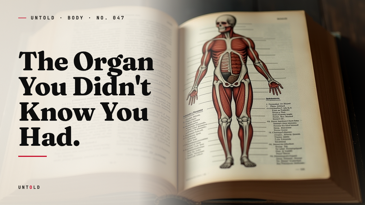
In 2017, a strange announcement made its way from a surgical journal into the popular press. Scientists, the headlines said, had discovered a new organ in the human body. It was a peculiar claim on its face. Organs are not the sort of thing that turn up unexpectedly. We have been dissecting ourselves for well over two thousand years, ever since the Greek physicians of Alexandria first opened the human abdomen. By the twenty-first century, the map of the body was supposed to be finished. There were no blank spaces left, no coastlines still uncharted.
And yet the announcement was, in a sense, true. What the researchers had done was not so much find something new as finally see something old correctly. The structure in question had been sitting inside every human being who ever lived. It had been sketched, sliced, described, and dismissed for centuries. The problem was not that anyone had missed it. The problem was that everyone had looked at it and drawn the wrong conclusion. This is the story of the mesentery, and of how a single stubborn surgeon in the west of Ireland persuaded medicine to rewrite one of its oldest textbooks.
The Forgotten Fold
Open the human abdomen and the first thing you notice is the intestine. Meters of it, coiled and looped, glistening and mobile. It looks, at first glance, like chaos. But intestine of that length cannot simply float free inside the belly. Something has to hold it, feed it, and keep it from twisting itself into a fatal knot. That something is the mesentery: a thin, fan-shaped sheet of tissue that fastens the gut to the back wall of the abdomen and carries the blood vessels, lymph nodes, and nerves the intestine depends on to survive.
For most of medical history, the mesentery was regarded as little more than upholstery. It was the packing tissue that kept the interesting organs in place. Anatomists paid it the attention one pays to bubble wrap. Worse, they got its basic architecture wrong. The prevailing view, cemented over the nineteenth century, held that the mesentery was not one thing at all. It came, the textbooks insisted, in fragments: a piece near the small intestine here, a separate scrap near the colon there, discontinuous structures with no clear relationship to one another. The word itself, from the Greek mesos (middle) and enteron (intestine), described a position rather than a coherent object.
This fragmented picture had the weight of authority behind it, but it was not the only picture ever drawn. Around 1508, Leonardo da Vinci filled his anatomical notebooks with studies of the human interior, and among them he rendered the mesentery as a single, continuous structure fanning out to support the gut. Leonardo, working from direct dissection with the eye of a painter rather than the assumptions of a physician, saw the tissue whole. He had no reason to break it into pieces because his hand simply followed what his eye reported.
But Leonardo’s notebooks were not published in his lifetime. They passed through private collections and lay unread for centuries, their insights sealed away while formal anatomy went on developing without them. By the time scholars pored over his drawings, the fragmented model was already orthodoxy. The organ nobody bothered to question had been correctly described five hundred years earlier, and then quietly forgotten.
How a Wrong Picture Got Written Into Stone
To understand why the error lasted so long, it helps to understand how anatomical knowledge is manufactured. Anatomy is not learned by staring at a whole body. It is learned in slices. A structure is understood by cutting through it at chosen planes and inferring the three-dimensional object from a series of two-dimensional cross sections. This works beautifully for compact, discrete organs like the kidney or the spleen. It works far less well for a thin, folded sheet that curves back on itself.
Slice a folded fan at an angle and you will see what appear to be several separate blades of material, each isolated in its own patch of the cut. Only by tracing the tissue continuously, following it around its curves rather than sampling it at intervals, does the illusion resolve into a single object. Generations of anatomists sliced the mesentery, saw the apparent gaps, and recorded fragments. Each teacher passed the fragmented model to the next generation, and each new dissection, interpreted through that inherited expectation, seemed to confirm it. The error was self-reinforcing. It was not merely written down; it was built into how students were trained to look.
The authority of the fragmented model was sealed by the most influential anatomy book ever printed. In 1858, the English surgeon Henry Gray published Gray’s Anatomy, illustrated by Henry Vandyke Carter, whose meticulous engravings shaped how generations of doctors visualized the human interior. The book became the standard by which all others were measured, reprinted and revised for over a century and a half. And Gray’s Anatomy, like everything else, described the mesentery as fragmented. When the definitive text says a thing, that thing is no longer a hypothesis. It is a fact taught to every medical student on earth, an assumption so foundational that questioning it feels less like science than like heresy.
A Surgeon Who Would Not Let It Go
The person who finally questioned it was J. Calvin Coffey, a colorectal surgeon at University Hospital Limerick in Ireland. Coffey’s specialty put him in an unusual position. Where an anatomist studies dead tissue sliced on a table, a surgeon works with the living structure, exposed and intact, day after day. Coffey spent his operating hours cutting near the mesentery to remove cancers of the bowel, and over years of doing so he developed the intuition that something in the textbooks did not match what lay under his hands.
The tissue did not behave like fragments. In the operating theatre, when he mobilized the intestine and traced the supporting membrane, it appeared continuous, a single sheet flowing without the breaks the diagrams promised. This is the kind of quiet discrepancy that most practitioners notice, shrug at, and forget. The textbook is the textbook; a busy clinician has patients to treat, not centuries of consensus to overturn. But Coffey did not forget it. He began, methodically, to collect evidence.
He and his team gathered specimens, more than a hundred over the course of their investigation, and examined the mesentery under the microscope, mapping its boundaries and tracing its edges. The pattern that emerged was consistent and unambiguous. The fragments that anatomists had described were an artifact of sectioning, an illusion produced by cutting a folded structure at unfortunate angles. Followed continuously, the tissue was one unbroken organ, folded back on itself like a closed fan. In every case they studied, the mesentery revealed itself as a single, continuous structure.
In 2012, Coffey published his early evidence for this continuity. The reception was cool. Centuries of teaching stood against him, and a claim this large, that one of the oldest facts in anatomy was simply wrong, invited skepticism rather than applause. Extraordinary claims demand extraordinary evidence, and the surgical world was not prepared to discard Gray’s Anatomy on the word of one team in Limerick. So Coffey did the only thing a scientist can do in the face of doubt. He gathered more. More dissections, more careful imaging, more painstaking documentation of the tissue’s true architecture.
The Textbook Rewritten
The turning point came in November 2016, when Coffey and his colleague Peter O’Leary published a review in The Lancet Gastroenterology and Hepatology laying out the case that the mesentery should be reclassified as a distinct, continuous organ 1. This time the argument carried. The evidence had accumulated to the point where the fragmented model was no longer defensible, and the prestige of the journal gave the claim a platform the earlier work had lacked.
The consequences were remarkable in their speed. Gray’s Anatomy itself, the very text that had enshrined the error, updated its pages to reflect the continuous structure. By 2017 the reclassification had filtered into medical curricula, and students began learning the mesentery as an organ rather than as scattered scraps of connective tissue. Coffey put the shift plainly: medicine was now saying it had an organ in the body that it had not counted before. What had changed was not the anatomy of any single human being. What had changed was the profession’s willingness to see the tissue as it had always been.
It is worth pausing on how modest the underlying discovery was. Nothing new was added to the body. No hidden chamber was opened, no unknown vessel traced. All that happened was that a structure long regarded as several things was recognized as one thing. And yet that reclassification mattered, because how we categorize the body determines what questions we are able to ask about it. A fragment is background. An organ demands a job description.
What the Mesentery Actually Does
Once the mesentery was granted the status of an organ, the obvious question followed: what is it for? The answers turn out to be more consequential than the old dismissal as packing tissue ever suggested.
The most basic function is structural. The mesentery tethers the intestines to the abdominal wall in an organized way, keeping meters of bowel from collapsing into a tangled, twisted heap. Without that anchoring, the gut could not maintain the arrangement it needs to move food along its length. Threaded through the tissue run the blood vessels that feed the intestine, the lymphatic channels that drain it, and the nerves that regulate it. In this sense the mesentery is not a passive scaffold but a highway, the route by which everything the gut requires is delivered and everything it discards is carried away.
But structure and transport may be the least interesting part of the story. The mesentery is dense with lymph nodes, and lymph nodes are stations of the immune system. This has led researchers to suspect that the organ plays a role in immune signaling, sampling the contents of the gut and relaying information about potential threats to the rest of the body. The intestine is one of the largest surfaces where the human body meets the outside world, a border patrolled constantly against invading microbes, and the mesentery sits precisely at that frontier.
The organ also stores fat, and here it may intersect with disease in ways only now coming into focus. In Crohn’s disease, a chronic inflammatory condition of the gut, the fat of the mesentery behaves abnormally, wrapping itself around inflamed segments of intestine in a phenomenon surgeons call creeping fat. For a long time this was viewed as a curiosity, a side effect of the disease rather than a participant in it. The reframing of the mesentery as an organ has encouraged a different reading: that the tissue may be an active player in the inflammation itself. Some surgeons have begun removing diseased mesentery along with the affected bowel in Crohn’s operations, and early observations suggest this may reduce the rate at which the disease returns 2. If that holds up, it would mean the mesentery is not merely a bystander in gut disease but part of its mechanism, and therefore part of its cure.
That possibility is why Coffey described the reclassification not as an endpoint but as a beginning. Once a structure is recognized as an organ with functions of its own, an entire field of inquiry opens around it: its role in health, its contribution to disease, the ways it might be targeted by new treatments. The mesentery had been sitting inside the abdomen the whole time, doing all of this work, while medicine filed it under miscellaneous.
The Body Still Keeps Secrets
There is a temptation to tell this story as a discovery, a tale of something found. But that framing misses what makes it strange. The mesentery was never lost. It was there in every dissection, drawn in every atlas, held in every pair of hands that ever operated on the human gut. Leonardo saw it correctly five centuries ago. What was missing was not the object but the perception. For over a hundred years the profession looked at a continuous organ and saw fragments, because that is what it had been trained to see.
That is the uncomfortable lesson buried in the anatomy. We had mapped the surfaces of distant planets and measured the light of galaxies billions of years old before we managed to see clearly a fan-shaped membrane inside our own bellies. Knowledge does not fail only when facts are absent. It fails when a wrong idea becomes so familiar that it stops being an idea and starts being the frame through which everything else is viewed. The fragmented mesentery was not a gap in the record. It was an error written so deeply into the record that correcting it required a surgeon willing to distrust the most authoritative book in his field.
So the next time your stomach turns, some quiet disturbance deep in the gut, it may be worth remembering the structure doing the holding. A single continuous fold, folded on itself, feeding and anchoring and quietly signaling, drawn wrong for a century and named at last only a few years ago. It held your insides together long before anyone bothered to see it whole.

Sources
- Coffey, J. C. and O’Leary, D. P., The mesentery: structure, function, and role in disease, The Lancet Gastroenterology and Hepatology, 2016. — https://www.thelancet.com/journals/langas/article/PIIS2468-1253(16)30026-7/fulltext
- Coffey, J. C. et al., Inclusion of the Mesentery in Ileocolic Resection for Crohn’s Disease is Associated with Reduced Surgical Recurrence, Journal of Crohn’s and Colitis, 2018. — https://academic.oup.com/ecco-jcc/article/12/10/1139/5001673
- Coffey, J. C. and Walsh, D., The mesentery: a reappraisal, University of Limerick / University Hospital Limerick research communications, 2016. — https://www.ul.ie/gems
- Gray, Henry (illustrations by Henry Vandyke Carter), Anatomy: Descriptive and Surgical (Gray’s Anatomy), first edition 1858. — https://en.wikipedia.org/wiki/Gray%27s_Anatomy
- Clerc, O. et al., Historical review of the mesentery and Leonardo da Vinci’s anatomical studies, Clinical Anatomy, 2017. — https://onlinelibrary.wiley.com/journal/10982353
- Kruepunga, N. et al., Anatomy of rotation and fixation of the human midgut and mesentery, Journal of Anatomy, 2018. — https://onlinelibrary.wiley.com/journal/14697580
- Byrne, J. et al., The mesentery: newly recognized organ and its clinical significance, Nature Reviews / Scientific American reporting, 2017. — https://www.scientificamerican.com/article/meet-your-interstitium-a-newfound-organ/
Related reading